Mbl2 Rat Monoclonal Antibody [Clone ID: 14D12]
Specifications
| Product Data | |
| Clone Name | 14D12 |
| Applications | ELISA, IF, IHC, WB |
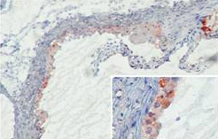
| Recommended Dilution | Immunohistochemistry on frozen sections (4): Cryostat sections (7µm) of mouse tissue were acetone fixed and stained o/n at 4°C in TBS/2% goat serum. (Ref.4). The typical starting working dilution is 1:10. It is recommended that solutions with a calcium concentration of 1 mM are used (14D12 is a calcium-dependent antibody). Immunoassays (1). Immunoflourescence (3,5): Hyphae of C. albicans were fixed on poly-l-lysin coated slides with ice-cold aceton. Slides were incubated for 1h at RT with 14D12. (Ref.3). Western blot (1,2): On a 12% non-reducing SDS-PAGE a prominent band of ca. 50Kda is detected. Under reducing conditions a 26 KDa band is detected. (Ref.2). The typical starting working dilution is 1:10. It is recommended that solutions with a calcium concentration of 1 mM are used (14D12 is a calcium-dependent antibody). Positive control: Mouse Serum and kidney tissue. |
| Reactivities | Mouse |
| Host | Rat |
| Isotype | IgG2a |
| Clonality | Monoclonal |
| Immunogen | Purified MBL-C |
| Specificity | This antibody detects MBL-C. |
| Formulation | PBS State: Purified State: Liquid 0.2 µm filtered Ig fraction Stabilizer: 0.1% bovine serum albumin Preservative: 0.02% sodium azide |
| Concentration | 0.1 mg/ml |
| Purification | Protein G |
| Database Link | |
| Background | Mannose binding lectin (MBL), also called mannose- or mannan-binding protein (MBP), is a member of the group of collectins. MBL is an important pattern-recognition receptor in the innate immune system. The protein mediates innate immune responses, such as activation of the complement lectin pathway and phagocytosis, to help fight infections. MBL is an oligomeric lectin that recognizes carbohydrates as mannose and N-acetylglucosamine on pathogens. MBL contains a cysteine rich, a collagen like and a carbohydrate recognition domain. Binding of MBL leads to the activation of MBL-associated serine proteases (MASP's). Activated MASP-2 cleaves C4 and C2 in a similar way as C1s do for the classical pathway (CP) leading to the formation of C4b2a, cleavage of the classical pathway convertase C3, and eventually complement activation up to the formation of the membrane attack complex. MBL is able to activate the complement pathway independent of the classical and alternative complement activation pathways. |
| Synonyms | MBP, Mannose-binding protein C, MBP-C, MBP1, Mannan-binding protein, Mannose-binding lectin, MBL2, MBL |
| Reference Data | |
Documents
| Product Manuals |
| FAQs |
| SDS |
{0} Product Review(s)
Be the first one to submit a review


 Germany
 Japan
Japan
 United Kingdom
 China
China


