LC3B (MAP1LC3B) Rabbit Polyclonal Antibody
Frequently bought together (1)
Other products for "MAP1LC3B"
Specifications
| Product Data | |
| Applications | IHC, WB |
| Recommended Dilution | Immunohistochemistry: 1:400, Immunocytochemistry/ Immunofluorescence: 1:75, Western Blot: 1:1000, Immunohistochemistry-Paraffin: 1:400 |
| Reactivities | Human, Mouse, Rat, Bovine, Porcine, Primate |
| Host | Rabbit |
| Clonality | Polyclonal |
| Immunogen | A synthetic peptide made to a C-terminal region of the human LC3 protein (within residues 50-120). [Swiss-Prot Q9H492] |
| Formulation | PBS with 30% Glycerol, 0.05% Sodium Azide. Store at 4C short term. Aliquot and store at -20C long term. Avoid freeze-thaw cycles. |
| Concentration | lot specific |
| Purification | Affinity purified |
| Conjugation | Unconjugated |
| Storage | Store at -20°C as received. |
| Stability | Stable for 12 months from date of receipt. |
| Gene Name | microtubule associated protein 1 light chain 3 beta |
| Database Link | |
| Background | Autophagy is a process of intracellular bulk degradation in which cytoplasmic components, including organelles, are sequestered within double-membrane vesicles that deliver the contents to the lysosome/vacuole for degradation. During macroautophagy, the sequestering vesicles, termed autophagosomes, fuse with the lysosome or vacuole resulting in the delivery of an inner vesicle (autophagic body) into the lumen of the degradative compartment. There are 16 proteins participating in the autophagy pathway in human. The autophagy protein LC3, a mammalian homologue of Atg8, was originally identified as microtubule-associated protein 1 light chain 3. It is a component of both the MAP1A and MAP1B microtubule-binding domains and the heavy-chain independent regulation of LC3 expression might modify MAP1 microtubule-binding activity during development. LC3 is the only known mammalian protein identified that stably associates with the autophagosome membranes. LC3-I is cytosolic and LC3-II is membrane bound and enriched in the autophagic vacuole fraction. The detection of LC3-I to LC3-II conversion is a useful and sensitive marker for distinguishing autophagy in mammalian cells. |
| Synonyms | 1BLC3; ATG8F; LC3B; MAP1A; MAP1LC3B-a |
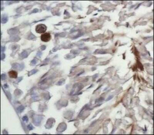
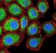
| Note | This LC3I antibody is useful for Western Blot, Immunocytochemistry/Immunofluorescence, and IHC-paraffin embedded sections. In Western Blot, a band is seen ~15kDa representing LC3I. In ICC/IF, observed staining showed inactivated LC3 throughout the cytoplasm of Neuro2a cells. In IHC-P, staining was observed in the cytoplasm of mouse testes tissue. Prior to immunostaining paraffin tissues, antigen retrieval with sodium citrate buffer (pH 6.0) is recommended. |
| Reference Data | |
Documents
| Product Manuals |
| FAQs |
| SDS |
{0} Product Review(s)
0 Product Review(s)
Submit review
Be the first one to submit a review
Product Citations
*Delivery time may vary from web posted schedule. Occasional delays may occur due to unforeseen
complexities in the preparation of your product. International customers may expect an additional 1-2 weeks
in shipping.


 Germany
 Japan
Japan
 United Kingdom
 China
China


