


 Islands")


")


")


")


")
")


 Germany
 Japan
Japan
 United Kingdom
 China
China
Primary Antibodies
Primary antibodies are Y-shaped glycoproteins that bind to a specific antigen with high specificity and affinity. The antigen can be a small molecule, peptide, or protein. Due to their high affinity, and high specificity, they are used in protein localization, purification, and quantification. Primary antibodies can be monoclonal or polyclonal depending on the epitopes they bind to; monoclonal antibodies bind to one specific epitope on antigens, whereas polyclonal antibodies bind to multiple different epitopes on the antigen.
OriGene offers monoclonal and polyclonal primary antibodies from various species with broad applications. Our antibodies are validated for applications like: IHC, ICC, ELISA, FACS, and WB. Over the last decade, we have developed our own branded antibodies, including TrueMAB?, UltraMAB?, and TrueRAB?. Our antibodies have been well-cited for their high specificity and affinity.
Featured products and services:


ABAT
ACTA2
ADIPOQ
AFP
AGR2

ALDH1L1
ALK
ALX4
AMACR

ANO1
AR
BBOX1
BCL2
BID
BMP4
BTLA
BUB1B
C14orf166
CA12
CD13
CD19
CD1C
CD2
CD274
CD38
CD3E
CD4
CD40
CD44
CD5


Popular marker citations
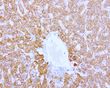
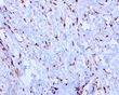
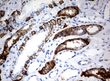
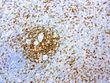
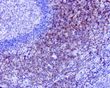
Anti-LGR5 Antibody
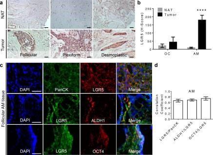
Title: LGR5 + epithelial tumor stem-like cells generate a 3D-organoid model for ameloblastoma.
PubMed ID: 32382005
Figure legend: "a Expression of?LGR5?in different histopathological types of AM ( n = 15). Scale bars, 20 ?m. NAT normal adjacent tissue (same patient). b The quantification of H-score of LGR5 expression in AM ( n = 15) and odontogenic cyst (OC) ( n = 6). H-Score of each sample was analyzed at least nine different areas by color deconvolution of ImageJ software and data are mean ? SD. Two-tailed unpaired Student?s t test. **** p < 0.0001. c Dual-color immunofluorescence study showed that about 66.3% of LGR5 signal was co-localized with the expression of pan-cytokeratin (Pan-CK) in the solid AM tissues ( n = 3). About 68.6% and 74.1% of LGR5 + cells co-expressed ALDH1 and OCT4, respectively. Scale bars, 20 ?m. The representative images are from the follicular AM. d The quantification of correlation coefficient of LGR5 and epithelial biomarker (Pan-CK) and stem cell-related genes (ALDH1 and OCT4) in the solid AMs ( n = 3). Each group was calculated at least three different areas by CellProfiler software and data are mean ? SD."
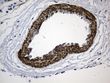
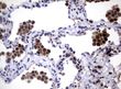
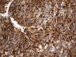
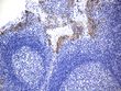
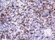
Anti- CD163 Antibody

Title: Collagen 1a1 Expression by Airway Macrophages Increases In Fibrotic ILDs and Is Associated With FVC Decline and Increased Mortality.
PubMed ID: 34867934
Figure legend: "COL1A1 is expressed by cells of myeloid origin and AMs. COL1A1 expression in fresh BAL cultures from a Non-IPF patient with CTD-ILD, stained with rabbit anti-human-COL1A1 and ToPro-633 nuclear stain and (A) mouse isotype control, (B) mouse anti-CD45 and (C) mouse?anti-CD163?antibodies. Arrows indicate CD45 and?CD163?positive cells expressing COL1A1. "?CellProfiler software and data are mean ? SD. "
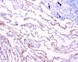
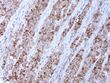
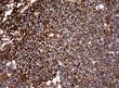
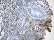
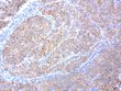
Anti- PD-L1 Antibody
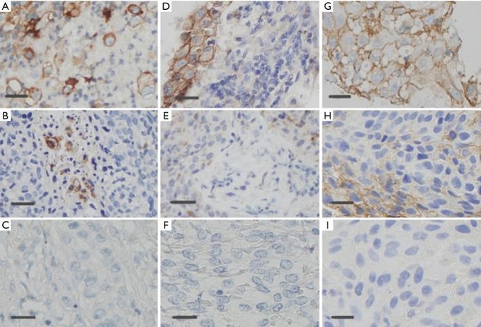
Title: Assessment of programmed cell death ligand-1 expression with multiple immunohistochemistry antibody clones in non-small cell lung cancer.
PubMed ID: 29607153
Figure legend: "Immunohistochemical staining of PD-L1 in NSCLCs, showing 100%, 30%, 0% PD-L1-positive tumor cells stained, respectively (400? magnification, bar =50 ?m). (A,B,C) SP263 assay demonstrating PD-L1 positive tumor cells; (D,E,F)?UMAB228?assay demonstrating PD-L1 positive tumor cells; (G,H,I) SP142 assay demonstrating PD-L1-positive tumor cells. NSCLCs, non-small cell lung cancer; PD-L1, programmed cell death ligand-1."
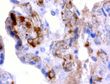
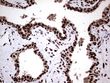
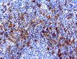
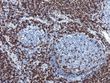
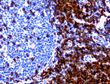
Anti- CD3E Antibody
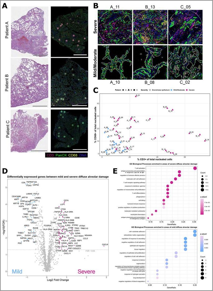
Title: Spatial transcriptomic characterization of COVID-19 pneumonitis identifies immune circuits related to tissue injury.
PubMed ID: 36472908
Figure legend: "Selection and annotation of areas of diffuse alveolar damage in COVID-19 lung tissues for transcriptomic analysis. ( A ) Merged immunofluorescence (IF) images of the lung samples from patients A, B and C (scale bars: 5mm, pan cytokeratin-green, DNA-blue, CD3-red, CD68-yellow). n=47 areas of interest (AOI) selected for transcript profiling are highlighted (blue circles-mild/moderate and magenta circles-severe pathology), of which n=46 passed quality control after sequencing. Labelled areas correspond to higher magnification examples in B . ( B ) Representative IF images of AOIs demonstrating the morphology and immune infiltrate observed within areas of severe and mild/moderate diffuse alveolar damage. AOIs spanned on average 0.2mm 2 (range: 0.05 to 0.33mm 2 ) with exclusion of empty space. ( C ) The proportion of CD3 + and CD68 + cells of total nucleated cells were derived from the immunofluorescent imaging, plotted for each AOI and coloured by histological severity (mild/moderate-blue, severe-magenta) or inclusion of bronchiolar epithelium (grey). AOI are annotated by patient: A ? circle, B ? triangle and C - square. ( D ) Differential gene expression between areas of severe and mild/moderate damage. Coloured and annotated genes have a fold change expression > 1.5 and a Benjamini-Hochberg (BH) adjusted p-value <0.05. ( E ) Selected GO biological processes significantly over-represented in genes differentially expressed between mild and severe areas of damage (BH corrected p < 0.05, one-sided Fisher?s exact test); see also Supplementary Table 3 for differentially expressed genes and all over-represented pathways. "


Our Antibodies
UltraMAB?

The Ultra Specific Antibodies Specificity tested by High-Density Protein Microarray Designed for anatomic pathology Focused on cancer research and diagnostics
TrueMAB?

Mouse Monoclonal Antibodies Full-length human proteins as antigens Recognize native epitopes of target proteins Extensive validations for WB, IHC, IF/ICC, IP, ELISA & Flow
TrueRAB?

Rabbit Monoclonal antibodies Higher sensitivity comparing to other species Smaller antigen for the accurate epitope mapping Extensive validations for WB, IHC, IF/ICC, IP, ELISA & Flow