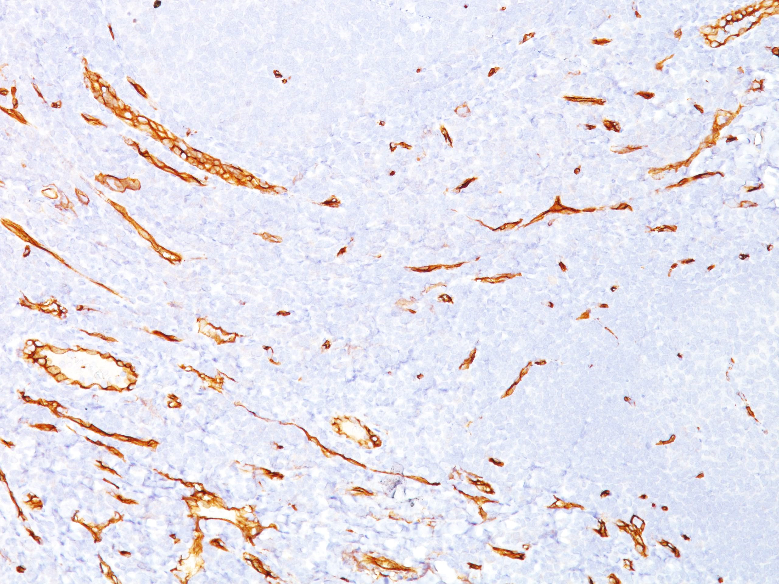
CD34 Mouse Monoclonal Antibody [Clone ID: SPM123]
Specifications
| Product Data | |
| Clone Name | SPM123 |
| Applications | FC, IF, IHC, IP, WB |
| Recommended Dilution | Flow Cytometry: 0.5-1 µg/million cells. Immunofluorescence: 0.5-1 µg/ml. Immunoprecipitation: 0.5-1 µg/500 µg protein. Western Blotting: 0.25-0.5 µg/ml. Immunohistochemistry on Frozen and Formalin-Fixed Paraffin Sections: 0.5-1 µg/ml for 30 min at RT. Staining of formalin-fixed tissues requires boiling tissue sections in 10mM Citrate Buffer, pH 6.0, for 10-20 min followed by cooling at RT for 20 minutes. Positive Control: KG-1 cells, Tonsil, or Angiosarcoma. |
| Reactivities | Human, Monkey |
| Host | Mouse |
| Isotype | IgG1 |
| Clonality | Monoclonal |
| Immunogen | Detergent solubilized vesicular suspension prepared from human term placenta. |
| Specificity | This Monoclonal Antibody recognizes a single chain, transmembrane, heavily glycosylated protein of 90-120kDa, which is identified as CD34. On the basis of differential sensitivity to degradation by specific enzymes, epitopes of monoclonal antibodies to CD34 are classified into three main categories, class I, class II and class III. It is a class II antibody whose epitope is resistant to neuraminidase but sensitive to glycoprotease and chymopapain. CD34 expression is a hallmark for identifying pluripotent hematopoietic stem or progenitor cells. Its expression is gradually lost as lineage committed progenitors differentiate. CD34 is a marker of choice for staining blasts in acute myeloid leukemia. In addition, CD34 is expressed by soft tissue tumors, such as solitary fibrous tumor and gastrointestinal stromal tumor. Its expression is also found in vascular endothelium. Additionally, it appears that proliferating endothelial cells express this molecule more than the non-proliferating endothelial cells. Anti-CD34 labels > 85% of angiosarcoma and Kaposi’s sarcoma, but with a lower specificity. Cellular Localization: Cell surface. Negative Species: Rat, Sheep, Cow and Dog. |
| Formulation | 10mM PBS State: Purified State: Liquid purified IgG fraction from Bioreactor Concentrate Stabilizer: 0.05% BSA Preservative: 0.05% Sodium Azide |
| Concentration | lot specific |
| Purification | Protein A/G Chromatography |
| Storage | Store undiluted at 2-8°C. |
| Stability | Shelf life: one year from despatch. |
| Predicted Protein Size | 90-110 kDa |
| Database Link | |
| Synonyms | Hematopoietic progenitor cell marker |
| Reference Data | |
Documents
| Product Manuals |
| FAQs |
{0} Product Review(s)
0 Product Review(s)
Submit review
Be the first one to submit a review
Product Citations
*Delivery time may vary from web posted schedule. Occasional delays may occur due to unforeseen
complexities in the preparation of your product. International customers may expect an additional 1-2 weeks
in shipping.


 Germany
 Japan
Japan
 United Kingdom
 China
China


