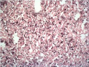
Adgre1 Rat Monoclonal Antibody [Clone ID: BM8]
Specifications
| Product Data | |
| Clone Name | BM8 |
| Applications | FC, IF, IHC, WB |
| Recommended Dilution | Immunohistochemistry on Frozen Sections (Ref.1,4): Tissue embedded in OCT Tissue Tec; fixed with acetone for 10 min at RT; incubation with 0.02 M sodium azide in PBS containing 0.1 % H2O2 for 10 min at RT to destroy endogenous peroxidase. Positive Control: spleen. Immunohistochemistry on Paraffin Sections (Rf.3): Fixation in 10% neutral buffered formalin for 24 h; blocking with non-immunized goat serum; microwaved for 6 min in citrate buffer. Positive Control: Splenic macrophages (Ref.3). Flow Cytometry (Ref.1,2): The recommended use of this reagent is 10 μl per 250.000 cells in a 100 μl total staining volume. The recommended use of this reagent is 10 μl per 250.000 cells in a 100 μl total staining volume. Western blot: Mouse bone-marrow derived macrophages; non-reduced; ~125 kDa (Ref.1); reduction with 2-mercaptoethanol destroys BM8 antigen. Positive Control: Mouse macrophages. Negative Control: Mouse fibroblasts or granulocytes. |
| Reactivities | Mouse |
| Host | Rat |
| Isotype | IgG2a |
| Clonality | Monoclonal |
| Specificity | The monoclonal antibody BM8 recognizes a 125 kDa extracellular macrophage membrane molecule, highly restricted to mature macrophage subpopulations residing in tissue. It is the only macrophage marker that is able to distinguish non-destructive from destructive inflammation processes in the pancreas. Furthermore it is a unique histological marker of the progression from peri-insulitis to beta-cell destruction and diabetes in a mouse diabetes model. It also detects Human heart macrophages. It does not detect Mouse granulocytes, Mouse mast cells, Mouse platelets, Mouse lymphocytes, Mouse fibroblasts, and Mouse endothelial cells. |
| Formulation | PBS Label: PE State: Liquid 0.2 µm filtered Ig fraction Stabilizer: 1% BSA Preservative: 0.02% Sodium Azide |
| Concentration | 0.1 mg/ml |
| Purification | Protein G Chromatography |
| Conjugation | PE |
| Database Link | |
| Background | This murine F4/80 glycoprotein contains seven-transmembrane (TM7) regions, which anchor the protein in the cell membrane, and thereby shows similarity in this region to G-protein-coupled receptors. The F4/80 molecule shares overall structural homology to other members of the epidermal growth factor (EGF)-TM7 family. The antigen is detected on tissue fixed macrophages in all organs tested so far (spleen, lymph nodes, thymus, liver, skin). It is also present on Langerhans cells in the skin and Kupffer cells in the liver. It is absent on granulocytes, lymphocytes and thrombocytes. The expression of F4/80 increases upon maturation of macrophage precursors in bone marrow and blood as well as in ontogeny. |
| Synonyms | Emr1, Gpf480 |
| Reference Data | |
Documents
| Product Manuals |
| FAQs |
| SDS |
{0} Product Review(s)
0 Product Review(s)
Submit review
Be the first one to submit a review
Product Citations
*Delivery time may vary from web posted schedule. Occasional delays may occur due to unforeseen
complexities in the preparation of your product. International customers may expect an additional 1-2 weeks
in shipping.


 Germany
 Japan
Japan
 United Kingdom
 China
China


