MHC Class II Ia (reacts with all haplotypes except H-2s) Mouse Polyclonal Antibody
CAT#: CL065F
MHC Class II Ia (reacts with all haplotypes except H-2s) mouse polyclonal antibody, FITC
Specifications
| Product Data | |
| Applications | Assay, IF |
| Recommended Dilution | Fluorescent Microscopy Analysis: 1/20. |
| Reactivities | Human, Mouse |
| Host | Mouse |
| Clonality | Polyclonal |
| Specificity | This anti-Mouse Ia alloantiserum is a broadly reactive Iak antiserum. The antiserum is prepared by immunizing A.TH mice with A.TL splenocytes. These two strains are congenic, differing at the I region of the H-2 complex but identical at the K and D regions. The two strains are characterized by the following H-2 haplotypes (Figure 1). The I region gene products (Ia antigens) potentially detected by this antiserum are those controlled by the following subregions: Ak, Bk, Ek, Ck i.e. it is a broadly reactive anti-Iak antiserum. It detects each of the following public and private specificities: Ia.1,2,3,7,15,19 and 22. As a result, this antiserum crossreacts with all standard haplotypes (i.e. H-2b,d,k,p,q,r) but does not crossreact with H-2s. The Ia antigens are expressed as cell surface antigens on lymphocyte subpopulations and macrophages and perhaps other non-lymphocytic cells. This fluorescein conjugated antibody can be used to localize or quantitate cells bearing the appropriate Ia antigens by fluorescence microscopy or cell sorter analysis. Since the A.TH anti-A.TL antiserum is cytotoxic, treatment of immunologically competent cell populations with this antiserum plus complement can quantitate cells bearing the corresponding Ia antigen or eliminate these cells from the population for functional studies. Ia Antigens on Human B Lymphocytes: A.TH anti-A.TL antiserum is strongly cytotoxic to human B lymphocytes but is not cytotoxic to human T-lymphocytes. This antiserum appears to recognize a determinant present on both mouse Ia and human HLA-DR antigens. The antiserum reacts with B lymphocytes of all individuals, although there is one report that B cells of some individuals are not reactive. In addition to human peripheral blood B lymphocytes, this antiserum reacts with chronic lymphatic leukemia (CLL) cells and at least a portion of monocytes. As a cytotoxic antibody, it can be used with complement for the enumeration or elimination of B cells, and is particularly well-suited as a positive control serum for B cell typing. The antiserum can also be used for biochemical studies of HLA-DR molecules. |
| Formulation | 0.05M carbonate buffer, pH 9.6, No preservatives added. Label: FITC State: Lyophilised Serum |
| Reconstitution Method | Restore with 0.5 ml of distilled water. |
| Purification | Protein G Chromatography |
| Conjugation | FITC |
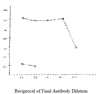
| Note | This antiserum is not sold as sterile. Filtration may result in severe loss of activity. If desired, sodium azide (0.02% final concentration) may be added as a preservative. Protocol: RECOMMENDED METHOD FOR STAINING MOUSE LYMPHOCYTES WITH FITC - ANTI-Ia ANTIBODY: Note: All procedures should be carried out on ice. 1. Prepare a cell suspension from the appropriate tissue in Dulbeccos phosphate buffered saline containing 0.3% bovine serum albumin plus 0.1% sodium azide (PBS-BSA). Remove red cells and dead cells (where necessary) by purification of viable lymphocytes on Lympholyte-M density cell separation medium. After washing, adjust the cell concentration to 2 x 10e7 cells/ml in PBS-BSA. 2. Add 50µl of cell suspension to the appropriate number of wells in a micro-titer round bottom plate. 3. Add 50 µl of appropriately diluted antibody (dilutions in PBS-BSA) to each well and mix. 4. Incubate for one hour on ice. 5. Pellet cells by centrifugation (500 x g for 3 minutes) and discard the supernatant. Wash x 2 with 100 µl of PBS-BSA. Remove supernatant. 6. For Fluorescent Microscopy: Resuspend in 50 µl of PBS-BSA. Place one drop on a microscope slide and cover with a cover glass. Score stained vs unstained cells in a fluorescent microscope. For Cell Sorter Analysis: Resuspend to the appropriate cell concentration and proceed as usual. Note: Prior to lyophilization, this product was clarified by centrigufation at 10000 x g for 10 minutes. If aggregates appear after prolonged storage of the reconstituted product, then centrifugation is recommended. SPECIAL NOTE: It is reported that azides react with lead and copper in plumbing to form compounds that may detonate on percussion. Please flush drains with large volumes of water when disposing of solutions containing sodium azide. LOT SPECIFICATIONS: Lot No: 510 Protein Concentration: 43.7 ± 4.9 mg/ml. Recommended Working Dilution (final): 1/20 Fluorescent Microscopy Analysis: Titration Method: As described above see Figure 2 Tissue Distribution: Method: As described above Cell Donor: A.TL Antibody Dilution Used: 1/20 Cell Source : % Fluorescent Stained Cells Enriched Splenic B Cells: 52% Lymph Node: 18% Thymus: 3% |
| Reference Data | |
Documents
| Product Manuals |
| FAQs |
| SDS |
{0} Product Review(s)
0 Product Review(s)
Submit review
Be the first one to submit a review
Product Citations
*Delivery time may vary from web posted schedule. Occasional delays may occur due to unforeseen
complexities in the preparation of your product. International customers may expect an additional 1-2 weeks
in shipping.


 Germany
 Japan
Japan
 United Kingdom
 China
China


