


 Islands")


")


")


")


")
")


 Germany
 Japan
Japan
 United Kingdom
 China
China
Endothelium Mouse Monoclonal Antibody [Clone ID: OX-43]
Product Images
Specifications
| Product Data | |
| Clone Name | OX-43 |
| Applications | FC |
| Recommended Dilution | Flow Cytometry. |
| Reactivities | Rat |
| Host | Mouse |
| Isotype | IgG1 |
| Clonality | Monoclonal |
| Immunogen | Rat Peritoneal Macrophages Immunocyte Donor: BALB/c Spleen Fusion Partner: NSO/U |
| Specificity | This monoclonal antibody recognizes a surface protein of MW 90 kDa and generally reacts with all vascular endothelium in the rat except that of brain capillaries. This is the reciprocal tissue pattern to that of the transferrin receptor. It has been shown that the expression of this Ab is on the luminal surface of blood vessels. This antibody labels all peritoneal macrophages, a sub-population of alveolar macrophages (65%) and rare interstitial cells in the brain and heart. In addition, anti-rat endothelium mAb labels circulating erythrocytes, 22% of peripheral blood mononuclear cells and 17% of nucleated cells in bone marrow. This antibody does not label granulocytes, dendritic cells, lymphocytes, or lymphocyte blasts, thymocytes, lymph node cells, mast cells and platelets. This antibody has been invaluable in the demonstration of molecular heterogeneity of vascular endothelium. |
| Formulation | PBS, 0.02% NaN3 and EIA grade BSA as a stabilizing protein to bring total protein concentration to 4-5 mg/ml Label: FITC State: Liquid purified |
| Concentration | 0.1 mg/ml |
| Purification | Protein G Chromatography |
| Conjugation | FITC |
| Background | The endothelium is located at the interface between the blood and the vessel wall. The cells are in close contact and form a slick layer that prevents blood cell interaction with the vessel wall as blood moves through the vessel lumen. The endothelium consists of simple squamous epithelium that lines the lumen of all blood vessels. It plays a critical role in the mechanics of blood flow, the regulation of coagulation, leukocyte adhesion, and vascular smooth muscle cell growth, and also serves as a barrier to the transvascular diffusion of liquids and solutes. For years the endothelium was thought of as an inert single layer of cells that passively allow the passage of water and other small molecules across the vessel wall. However, this dynamic tissue performs many other active functions, such as the secretion and modification of vasoactive substances and the contraction and relaxation of vascular smooth muscle. |
| Synonyms | endothelial cells, endothelial marker |
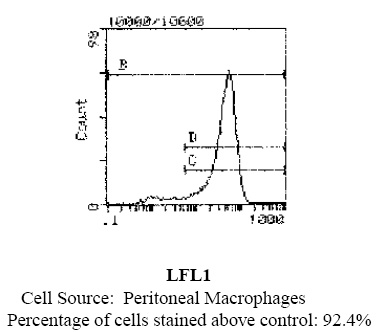
| Note | Protocol: FLOW CYTOMETRY ANALYSIS: Method: 1. Prepare a cell suspension in media A. For cell preparations, deplete the red blood cell population with Lympholyte®-Rat cell separation medium. 2. Wash 2 times. 3. Resuspend the cells to a concentration of 2x10e7 cells/ml in media A. Add 50µl of this suspension to each tube (each tube will then contain 1 x 10e6 cells, representing 1 test). 4. To each tube, add 0.2-0.5 µg* of this Ab. 5. Vortex the tubes to ensure thorough mixing of antibody and cells. 6. Incubate the tubes for 30 minutes at 4°C. (It is recommended that the tubes are protected from light, since most fluorochromes are light sensitive.) 7. Wash 2 times at 4°C. 8. Resuspend the cell pellet in 50 µl ice cold media B. 9. Transfer to suitable tubes for flow cytometric analysis containing 15 µl of propidium iodide at 0.5 mg/ml in PBS. This stains dead cells by intercalating in DNA. Media: A. Phosphate buffered saline (pH 7.2) + 5% normal serum of host species + sodium azide (100 µl of 2M sodium azide in 100 mls). B. Phosphate buffered saline (pH 7.2) + 0.5% Bovine serum albumin + sodium azide (100 µl of 2M sodium azide in 100 mls). Results - Tissue Distribution: Rat Strain: Fischer Cell Concentration: 1x10e6 cells per test Antibody Concentration Used: 0.5 µg/10e6 cells Isotypic Control: FITC Mouse IgG1,k Cell Source Percentage of cells stained above control: Thymus: 3.0% Peritoneal Macrophages: 92.4% Results - Strain Distribution: Antibody Concentration Used: 0.5 mg/10e6 cells Strains Tested: Wistar, Brown Norway, Buffalo, Fischer 344 Positive: Buffalo, Brown Norway, Fischer 344, Wistar Negative: none |
| Reference Data | |
Documents
| Product Manuals |
| FAQs |
| SDS |
{0} Product Review(s)
0 Product Review(s)
Submit review
Be the first one to submit a review


Product Citations
*Delivery time may vary from web posted schedule. Occasional delays may occur due to unforeseen
complexities in the preparation of your product. International customers may expect an additional 1-2 weeks
in shipping.