


 Islands")


")


")


")


")
")


 Germany
 Japan
Japan
 United Kingdom
 China
China
CD11b Mouse Monoclonal Antibody [Clone ID: OX-42]
Product Images
Specifications
| Product Data | |
| Clone Name | OX-42 |
| Applications | FC, IHC |
| Recommended Dilution | Flow cytometry. Immunohistochemistry on frozen and paraffin sections. |
| Reactivities | Rat |
| Host | Mouse |
| Isotype | IgG2a |
| Clonality | Monoclonal |
| Immunogen | Rat Peritoneal Macrophages Immunocyte Donor: BALB/c spleen Fusion Partner: NSO/U mouse myeloma cell line |
| Specificity | This monoclonal antibody recognizes most macrophages including resident peritoneal macrophages, kupffer cells, but only approximately 35% of alveolor macrophages (2). CL042B also labels dendritic cells extensively, granulocytes and cells with morphology of microglia in the brain. It precipitates three polypeptides of M.W. 160, 103 and 95 kDa. This antibody inhibits complement mediated rosettes and is probably the rat equivalent of the human receptor of iC3b called CR3. This anti-rat CD11b/c mAb should prove useful in the recognition of macrophages and microglia. |
| Formulation | PBS, 0.02% NaN3 and EIA grade BSA was added as a stabilizing protein to bring total protein concentration to 4-5 mg/ml. Label: Biotin State: Liquid purified Ig |
| Concentration | 0.1 mg/ml |
| Purification | Protein G Chromatography |
| Conjugation | Biotin |
| Background | CD11b is implicated in various adhesive interactions of monocytes, macrophages and granulocytes as well as in mediating the uptake of complement coated particles. It is identical to CR3, the receptor for the iC3b fragment of the third complement component. It probably recognizes the RGD peptide in C3b. CD11b is also a receptor for fibrinogen, factor X and ICAM1. It recognizes P1 and P2 peptides of fibrinogen gamma chain. The Mac1 CD11b antigen is present on macrophages, granulocytes, natural killer cells, blood monocytes. CD11b is expressed on 8% spleen cells, 44% bone marrow cells and less than 1% of thymocytes and is commonly used as a microglial marker in nervous tissue. |
| Synonyms | ITGAM, CR3A, CR-3 alpha chain, Integrin alpha-M, MAC1 |
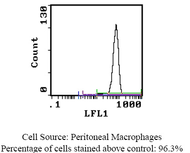
| Note | Protocol: FLOW CYTOMETRY ANALYSIS: Method: 1. Prepare a cell suspension in media A. For cell preparations, deplete the red blood cell population with Lympholyte®-Rat cell separation medium. 2. Wash 2 times. 3. Resuspend the cells to a concentration of 2x10e7 cells/ml in media A. Add 50 µl of this suspension to each tube (each tube will then contain 1 x 10e6 cells, representing 1 test). 4. To each tube, add 0.2-0.1 µg* of this Ab per 10e6 cells. 5. Vortex the tubes to ensure thorough mixing of antibody and cells. 6. Incubate the tubes for 30 minutes at 4°C. 7. Wash 2 times at 4°C. 8. Add 100 µl of secondary antibody (Streptavidin-FITC) at a 1:500 dilution. 9. Incubate tubes at 4°C for 30 - 60 minutes (It is recommended that tubes are protected from light since most fluorochromes are light sensitive). 10. Wash 2 times at 4°C. 11. Resuspend the cell pellet in 50 µl ice cold media B. 12. Transfer to suitable tubes for flow cytometric analysis containing 15 µl of propidium iodide at 0.5 mg/ml in PBS. This stains dead cells by intercalating in DNA. Media: A. Phosphate buffered saline (pH 7.2) + 5% normal serum of host species + sodium azide (100 µl of 2M sodium azide in 100 mls). B. Phosphate buffered saline (pH 7.2) + 0.5% Bovine serum albumin + sodium azide (100 µl of 2M sodium azide in 100 mls). Results - Tissue Distribution by Flow Cytometry Analysis: Rat Strain: Buffalo Cell Concentration: 1x10e6 cells per test Antibody Concentration Used: 0.1 µg/10e6 cells Cell Source: Peritoneal Macrophages Isotypic Control: Biotin Mouse IgG2a, k Cell Source Percentage of cells stained above control: Peritoneal Macrophages: 96.3% Thymus: 0.8% STRAIN DISTRIBUTION: Antibody Concentration: 0.5 µg/10e6 cells Strains Tested: Wistar, Buffalo, Brown Norway, Fischer 344 Positive: Wistar, Buffalo, Brown Norway, Fischer 344 Negative: None |
| Reference Data | |
Documents
| Product Manuals |
| FAQs |
| SDS |
{0} Product Review(s)
0 Product Review(s)
Submit review
Be the first one to submit a review


Product Citations
*Delivery time may vary from web posted schedule. Occasional delays may occur due to unforeseen
complexities in the preparation of your product. International customers may expect an additional 1-2 weeks
in shipping.