PIP5K3 (PIKFYVE) Human Gene Knockout Kit (CRISPR)
CAT#: KN412288
PIKFYVE - KN2.0, Human gene knockout kit via CRISPR, non-homology mediated.
KN2.0 knockout kit validation
KN412288 is the updated version of KN212288.
USD 1,290.00
2 Weeks*
Size
Other products for "PIKFYVE"
Specifications
| Product Data | |
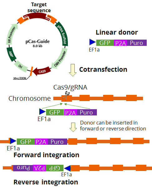
| Format | 2 gRNA vectors, 1 linear donor |
| Donor DNA | EF1a-GFP-P2A-Puro |
| Symbol | PIKFYVE |
| Locus ID | 200576 |
| Disclaimer | The kit is designed based on the best knowledge of CRISPR technology. The system has been functionally validated for knocking-in the cassette downstream the native promoter. The efficiency of the knock-out varies due to the nature of the biology and the complexity of the experimental process. |
| Reference Data | |
| RefSeq | NM_001002881, NM_001178000, NM_015040, NM_152671 |
| Synonyms | CFD; KIAA0981; MGC40423; PIKfyve; PIP5K |
| Summary | Phosphorylated derivatives of phosphatidylinositol (PtdIns) regulate cytoskeletal functions, membrane trafficking, and receptor signaling by recruiting protein complexes to cell- and endosomal-membranes. Humans have multiple PtdIns proteins that differ by the degree and position of phosphorylation of the inositol ring. This gene encodes an enzyme (PIKfyve; also known as phosphatidylinositol-3-phosphate 5-kinase type III or PIPKIII) that phosphorylates the D-5 position in PtdIns and phosphatidylinositol-3-phosphate (PtdIns3P) to make PtdIns5P and PtdIns(3,5)biphosphate. The D-5 position also can be phosphorylated by type I PtdIns4P-5-kinases (PIP5Ks) that are encoded by distinct genes and preferentially phosphorylate D-4 phosphorylated PtdIns. In contrast, PIKfyve preferentially phosphorylates D-3 phosphorylated PtdIns. In addition to being a lipid kinase, PIKfyve also has protein kinase activity. PIKfyve regulates endomembrane homeostasis and plays a role in the biogenesis of endosome carrier vesicles from early endosomes. Mutations in this gene cause corneal fleck dystrophy (CFD); an autosomal dominant disorder characterized by numerous small white flecks present in all layers of the corneal stroma. Histologically, these flecks appear to be keratocytes distended with lipid and mucopolysaccharide filled intracytoplasmic vacuoles. Alternative splicing results in multiple transcript variants encoding distinct isoforms. [provided by RefSeq, May 2010] |
Documents
| Product Manuals |
| FAQs |
| SDS |
Resources
Other Versions
| SKU | Description | Size | Price |
|---|---|---|---|
| GA116355 | PIKFYVE CRISPRa kit - CRISPR gene activation of human phosphoinositide kinase, FYVE-type zinc finger containing |
USD 1,290.00 |
{0} Product Review(s)
0 Product Review(s)
Submit review
Be the first one to submit a review
Product Citations
*Delivery time may vary from web posted schedule. Occasional delays may occur due to unforeseen
complexities in the preparation of your product. International customers may expect an additional 1-2 weeks
in shipping.


 Germany
 Japan
Japan
 United Kingdom
 China
China
