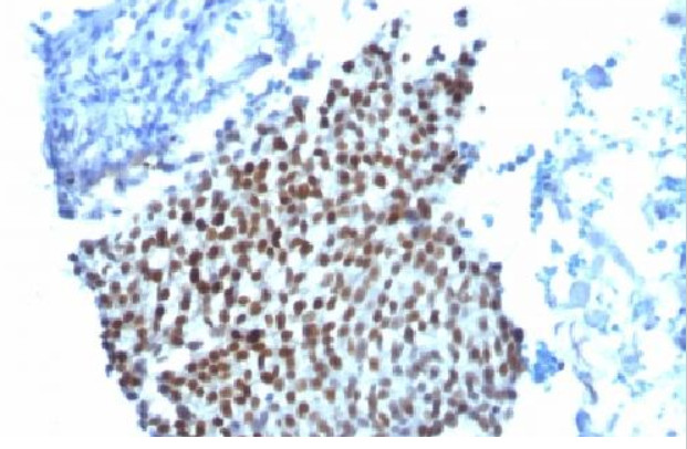
p21 (CDKN1A) Mouse Monoclonal Antibody [Clone ID: WA-1]
Specifications
| Product Data | |
| Clone Name | WA-1 |
| Applications | FC, IF, IHC |
| Recommended Dilution | Flow Cytometry: 0.5-1 μg/106 cells in 100 µl. Immunofluorescence: 1-2 μg/ml. Immunohistochemistry on Paraffin Sections: 0.5-1 µg/ml for 30 minutes at RT. Staining of formalin-fixed tissues requires boiling tissue sections in 10mM citrate buffer, pH 6.0, for 10-20 min followed by cooling at RT for 20 minutes. Positive Control: HeLa Cells. Skin, colon, or breast carcinoma. |
| Reactivities | Chimpanzee, Human, Monkey, Mouse, Rat |
| Host | Mouse |
| Isotype | IgG1 |
| Clonality | Monoclonal |
| Immunogen | Recombinant Human p21 protein. |
| Specificity | This Monoclonal antibody WA-1 (same as HJ21) recognizes a 21kDa protein, identified as the p21WAF1 tumor suppressor protein. This Monoclonal antibody WA-1 is highly specific to p21 and shows no cross-reaction with other closely related mitotic inhibitors. It is interesting to note that p21 expression is induced by wild type, but not mutant p53. The HJ21/WA-1 antibody clone has been used to demonstrate this phenomenon by western blot (Blaydes, 2001). The inability of mutant p53 to induce p21 essentially means that normal functions of p21 are compromised when p53 gene mutations are present. Since p53 gene mutations are present in up to 50% of human cancers, the loss of p21 function in cancer is significant. In its normal functioning state, p21 binds to cyclin/CDK complexes. When it binds to these complexes, it inhibits their kinase activity which, in turn, stops cell cycle progression and hence p21 gains its reputation as a mitotic cell cycle inhibitor. In this regard, the antibody showed that decreased Cdk2-cyclin E1 activity corresponded with a decrease in cyclin E1 and increase in p21 protein levels (White, 2005). The antibody is highly specific to p21; it does not cross-react with other closely related mitotic inhibitors. Specificity validations include antibody recognition of recombinant p21 by western blot (Koike, 2011) and in direct ELISA assays (Rossner, 2002 & 2007). Although the exact epitope for the antibody has not been mapped, the epitope appears to be different from those recognize by other p21 antibody clones. |
| Formulation | 10mM PBS State: Purified State: Liquid purified IgG fraction from Bioreactor Concentrate Stabilizer: 0.05% BSA Preservative: 0.05% Sodium Azide |
| Concentration | lot specific |
| Purification | Protein A/G Chromatography |
| Storage | Store undiluted at 2-8°C. DO NOT FREEZE! |
| Stability | Shelf life: one year from despatch. |
| Predicted Protein Size | 21 kDa |
| Database Link | |
| Background | p21 (WAF1), a member of the cycle dependent kinase (CDK) inhibitor family, was first identified as a tumor suppressor or anti-oncogenic protein and negative regulator of the cell cycle. Over the years it became apparent that p21 has broad functions in regulating fundamental cellular programs including proliferation, differentiation, migration, senescence, and has both anti-oncogenic and oncogenic properties (reviewed in Romanov, 2012). Many antibody and other studies have demonstrated that p21 levels are frequently elevated in situations associated with reduced proliferation, including differentiation and senescence, and decreased in proliferative states. Subcellular localization may significant and nuclear antibody staining may be indicative of p21 functioning as a cell cycle inhibitor, tumor suppressor, or in a senescence program. In contrast, cytoplasmic antibody staining may indicate p21 is acting an oncogene through regulatation of migration, proliferation, or apoptosis. |
| Synonyms | CAP20, CDKN1, CIP1, MDA6, MDA-6, PIC1, SDI1, WAF1 |
| Reference Data | |
Documents
| Product Manuals |
| FAQs |
{0} Product Review(s)
0 Product Review(s)
Submit review
Be the first one to submit a review
Product Citations
*Delivery time may vary from web posted schedule. Occasional delays may occur due to unforeseen
complexities in the preparation of your product. International customers may expect an additional 1-2 weeks
in shipping.


 Germany
 Japan
Japan
 United Kingdom
 China
China


