VE Cadherin (CDH5) (Extracell. Dom) Mouse Monoclonal Antibody [Clone ID: BV9]
CAT#: AM26286FC-N
VE Cadherin (CDH5) (Extracell. Dom) mouse monoclonal antibody, clone BV9, FITC
Specifications
| Product Data | |
| Clone Name | BV9 |
| Applications | ELISA, FC, FN, IF, IHC, IP, WB |
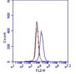
| Recommended Dilution | Immunohistochemistry on frozen sections (2): Acetone fixed sections were blocked with horse serum and incubated with antibody BV9 for 30 minutes (Ref.2). The typical starting working dilution is 1:10. Flow Cytometry (4): Antibody BV9 stains the extracellular domain of VE-cadherin. As negative control an IgG isotype control was used (Ref.4). The typical starting working dilution is 1:10. Functional Assays (5-8): Antibody BV9 functions as an antagonist. The antibody was functionally tested by adding 10-50µg/ml antibody BV9 to cell culture. The antibody blocks VE-cadherin causing a redistribution of VE-cadherin away from intracellular junctions(Ref.5, 6). Immunoassays: Antibody BV9 can function as coat and detector. Immunoprecipitation (3). Western Blot (1,3): A reduced sample treatment and 7.5% SDS-Page was used. The band size is 130-140kDa (Ref.3). The typical starting working dilution is 1:10. Immunoflourescence (3,5,8): Cells on coverslips were fixed with 3% paraformaldehyde and permeabilized with 0.5% Triton X-100 before incubation with antibody BV9 (Ref.5, 8). Positive Control: HUVECs grown on coverslips. |
| Reactivities | Human |
| Host | Mouse |
| Isotype | IgG2a |
| Clonality | Monoclonal |
| Specificity | The monoclonal antibody BV9 binds to the extracellular domain (EC3-EC4) of human VE-cadherin (vascular endothelial cadherin). |
| Formulation | PBS Label: FITC State: Liquid 0.2 µm filtered Ig fraction Stabilizer: 1% BSA Preservative: 0.02% Sodium Azide |
| Concentration | lot specific |
| Purification | Protein G Chromatography |
| Conjugation | FITC |
| Storage | Store undiluted at 2-8°C. DO NOT FREEZE! |
| Stability | Shelf life: one year from despatch. |
| Gene Name | cadherin 5 |
| Database Link | |
| Background | Endothelial cells control the passage of plasma constituents and circulating cells from blood to the underlying tissues. VE-cadherin is of vital importance for the maintenance and control of endothelial cell contacts. Mechanisms that regulate VE-cadherin–mediated adhesion are important for the control of vascular permeability and leukocyte extravasation. VE-cadherin regulates various cellular processes such as cell proliferation and apoptosis and modulates vascular endothelial growth factor receptor functions. Therefore, VE-cadherin is also essential during embryonic angiogenesis. The specialized function of VE-cadherin is lost or impaired in several pathological conditions - including inflammation, sepsis, ischemia and diabetes - which leads to severe, and sometimes fatal, organ dysfunction. Furthermore, abnormal increase in vascular permeability is often observed in pathological conditions, such as tumor-induced angiogenesis, macular degeneration, allergy, and brain stroke. |
| Synonyms | VE-Cadherin, Vascular endothelial cadherin, CDH5 |
| Reference Data | |
Documents
| Product Manuals |
| FAQs |
{0} Product Review(s)
Be the first one to submit a review


 Germany
 Japan
Japan
 United Kingdom
 China
China


