CD31 (PECAM1) Mouse Monoclonal Antibody [Clone ID: BC16-6.4EF]
Specifications
| Product Data | |
| Clone Name | BC16-6.4EF |
| Applications | FC, FN, IF, IHC, WB |
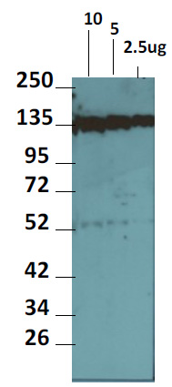
| Recommended Dilution | Western Blot: Use the BC16-6.4EF antibody at 2-3 µg/ml in Tris buffered Saline with 0.05% Tween 20 and 5% non-fat dry milk (Blotto) or similar diluents. The antibody reacts with a band of Approximate Molecular size 135kDa. Immunostaining: Use the BC16-6.4EF antibody at 2-3 µg/ml diluted with PBS containing 1% BSA. Indirect Immunofluorescence. Immunohistochemistry: 3-5 µg/ml. The BC16-6.4EF antibody may be used on endothelial cells such as HUVEC cells grown on chamber slides, cytospins and cryosections of Human tissue. For staining, the cultured cells and cryosections should be fixed in 1-2% paraformaldehyde for 30 mins, permeabilized in 0.25% Triton X 100 in PBS for 30 mins and non-specific binding blocked with 1% BSA in PBS. The primary antibody may be diluted in PBS with 1% BSA and incubated on cells/tissue overnight at 4°C. The antibody may also be used to inhibit endothelial cell–cell interactions. Suggested Positive Control Cells and Tissues: For staining and Western blotting, Human Umbilical Endothelial Cells (Thermo Fisher). For immunohistochemical staining, cryosections of most Human tissue with vasculature may be used. Immunohistochemistry on Paraffin Sections: 2-5 µg/ml. Heat induced antigen retrieval with citrat buffer, pH 6.2 using a pressure cooker was preformed. Sections were blocking using a commercially available casein solution. Signal was generated using a commercially available polymer HRP detection system and DAB. |
| Reactivities | Human, Mouse, Rat |
| Host | Mouse |
| Isotype | IgG1 |
| Clonality | Monoclonal |
| Immunogen | Biochemically characterized crude subcellular/membrane fraction obtained from HUVEC cells. |
| Specificity | The antibody was tested against normal and Human tumor tissue and HUVEC cells. The antibody was also tested against Mouse and Rat tissues. Staining of vasculature is observed in all tissues examined. The HUVEC cells showed cell surface and cytoplasmic staining. |
| Formulation | PBS, pH 7.2 State: Purified State: Liquid purified IgG fraction from Tissue Culture Supernatant Preservative: 0.05% Sodium Azide |
| Concentration | lot specific |
| Purification | Protein G Sepharose Chromatography |
| Storage | Store undiluted at 2-8°C for one month or (in aliquots) at -20°C for longer. Avoid repeated freezing and thawing. |
| Stability | Shelf life: one year from despatch. |
| Predicted Protein Size | Approximately 81 kDa (Predicted), a higher band of about 135kDa is observed. The higher Molecular Weight observed may be due post translational modifications including glycosylation. A significantly weaker band of Approximately 55kDa is also observed. |
| Gene Name | platelet and endothelial cell adhesion molecule 1 |
| Database Link | |
| Background | Platelet endothelial cell adhesion molecule-1 (PECAM-1) or cluster designation 31 (CD31) is a member of the immunoglobulin (Ig) gene superfamily and a transmembrane glycoprotein. The CD31 protein is widely expressed by cells of the vascular endothelium lineage, platelets, Kupffer cells, granulocytes, megakaryocytes, monocytes, neutrophils, and some types of T-cells. CD31 functions as a cell adhesion molecule in endothelial cell homotypic interactions in the process of angiogenesis. CD31 is also involved in leukocyte migration and integrin activation. It plays a key role in the adhesion cascade between the endothelial cells and inflammatory cells, enabling leukocyte migration in inflammatory sites. CD31 is one of the best markers for benign and malignant vascular tumors. It is expressed in certain tumors including angiomas and angiosarcomas including epitheloid hemangioendothelioma, epitheloid sarcoma-like hemangioendothelioma. Its expression in Kaposi sarcoma lesions is variable. Human CD31 comprises 738 amino acids and its predicted molecular weight would therefore be approximately 81kDa. However the observed molecular weight, as reported by various research groups including our own, is higher and at around 120kDa and 140kDa. The disparity probably reflects post translational modifications including glycosylation and probably because different isoforms of the protein may exist. |
| Synonyms | PECAM-1, EndoCAM, GPIIA' |
| Reference Data | |
Documents
| Product Manuals |
| FAQs |
{0} Product Review(s)
0 Product Review(s)
Submit review
Be the first one to submit a review
Product Citations
*Delivery time may vary from web posted schedule. Occasional delays may occur due to unforeseen
complexities in the preparation of your product. International customers may expect an additional 1-2 weeks
in shipping.


 Germany
 Japan
Japan
 United Kingdom
 China
China


