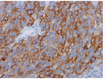
MelanA (MLANA) Mouse Monoclonal Antibody [Clone ID: M2-7C10]
Specifications
| Product Data | |
| Clone Name | M2-7C10 |
| Applications | FC, IF, IHC, IP, WB |
| Recommended Dilution | ELISA: Use Antibody without BSA for Coating. Western Blot: 0.5-1 µg/ml. Immunoprecipitaion: 1-2 µg/500 µg protein lysate. Flow Cytometry: 0.5-1 µg/106 cells. Immunofluorescence: 1-2 µg/ml. Immunohistochemistry on Frozen Sections. Immunohistochemistry on Paraffin Sections: 0.5-1 µg/ml for 30 minutes at RT. Staining of formalin-fixed tissues is enhanced by boiling tissue sections in 10 mM citrate buffer, pH 6.0 for 10-20 min followed by cooling at RT for 20 min. Positive Control: SK-MEL-13 and SK-MEL-19 Melanoma cell lines; Melanomas. |
| Reactivities | Human |
| Host | Mouse |
| Isotype | IgG2b |
| Clonality | Monoclonal |
| Immunogen | Recombinant Human MART-1 protein. |
| Specificity | This MART-1 antibody clone M2-7C10 recognizes a protein doublet of 20-22kDa, identified as MART-1 (Melanoma Antigen Recognized by T cells 1) or Melan-A. This MART-1 antibody clone M2-7C10 does not react with Mouse or Rat. Others species not tested. Researchers should use the MART-1 Antibody clone M2-9E3 (Cat.-No AM32813PU), which also recognizes Human MART-1, to detect Mouse or Rat MART-1. The clone M2-7C10 MART-1 antibody labels melanomas and other tumors showing melanocyte differentiation, and is widely used for assessing melanomas (Campoli, 2012, Ohsie, 2012, Collins, 2012). Analysis of melanoma lesions with this antibody shows that there is significant heterogeneity of expression of MART-1 both as a percentage of cells and in intensity of expression (Marincola, 1996). The reactivity of the MART-1 antibody is not restricted to melanoma, and the antibody has also been shown to label some mesenchymal tumors and sarcomas (Campoli, 2012). The exact eptiope recognized by this MART-1 antibody has not been mapped. However, the MART-1 epitope recognized by this antibody appears to be different from that recognized by the MART-1 antibody clone M2-9E3 (Cat.-No AM32813PU) (Kawakami, 1997). Researchers often use more than one antibody against a given specificity to help follow up and validate results. Hence, it may be useful to use both the Cat.-No AM32812PU and Cat.-No AM32813PU antibodies in parallel to obtain additional information about MART-1 expression. Cellular Localization: Cytoplasmic. |
| Formulation | 10mM PBS State: Purified State: Liquid purified IgG fraction from Bioreactor Concentrate Stabilizer: 0.05% BSA Preservative: 0.05% Sodium Azide |
| Concentration | lot specific |
| Purification | Affinity Chromatography on Protein A/G |
| Storage | Store undiluted at 2-8°C. |
| Stability | Shelf life: one year from despatch. |
| Predicted Protein Size | 20-22 kDa (Doublet) |
| Database Link | |
| Background | MART-1 (Melanoma Antigen Recognize by T cells 1), also known as Melan-A, is an 18 kDa melanocyte differentiation antigen recognized by autologous cytotoxic T lymphocytes. MART-1 is expressed in melanosomes and the endoplasmic reticulum. MART-1 is the most widely used marker for identifying malignant melanoma, the most deadly form of skin cancer, and facilitating complete removal of the primary tumor (Campoli, 2012). In this regard, MART-1 is used both as a confirmatory marker for melanocyte differentiation in S100 (protein present in melanocytes) positive lesions and a primary marker to evaluate the extent of melanocyte tumors (Ohsie, 2012, Collins, 2012). MART-1 specific monoclonal antibodies have high sensitivity (75-92%) and specificity (95-100%) for melanoma (Campoli, 2012, Oshie et.al, 2012). |
| Synonyms | Melan-A protein, MLANA, Antigen SK29-AA, LB39-AA, MelanA, MART1 |
| Reference Data | |
Documents
| Product Manuals |
| FAQs |
{0} Product Review(s)
0 Product Review(s)
Submit review
Be the first one to submit a review
Product Citations
*Delivery time may vary from web posted schedule. Occasional delays may occur due to unforeseen
complexities in the preparation of your product. International customers may expect an additional 1-2 weeks
in shipping.


 Germany
 Japan
Japan
 United Kingdom
 China
China


