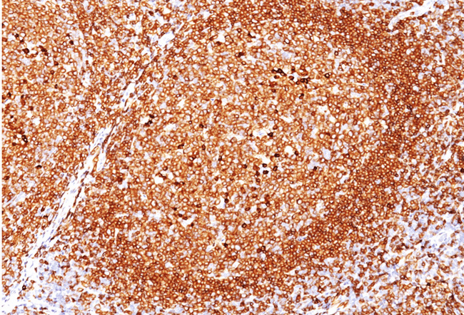
CD79A Mouse Monoclonal Antibody [Clone ID: JCB117]
Specifications
| Product Data | |
| Clone Name | JCB117 |
| Applications | FC, IF, IHC, IP, WB |
| Recommended Dilution | ELISA: Use BSA free Antibody for coating. Flow Cytometry: 0.5-1 µg/million cells. Immunofluorescence: 0.5-1 µg/ml. Western Blotting: 0.5-1 µg/ml. Immunoprecipitation: 0.5-1 µg/500 µg protein lysate. Immunohistochemistry on Frozen and Formalin-Fixed Paraffin Sections: 0.5-1 µ/ml for 30 minutes at RT. Staining of formalin-fixed tissues requires boiling tissue sections in 10mM citrate buffer, pH 6.0, for 10-20 min followed by cooling at RT for 20 minutes. Positive Control: Daudi or Ramos cells. Germinal center B-cells in a lymph node or tonsil. |
| Reactivities | Human |
| Host | Mouse |
| Isotype | IgG1 |
| Clonality | Monoclonal |
| Immunogen | A synthetic peptide corresponding to aa 202-216 (GTYQDVGSLNIADVQ) of Human CD79a protein. |
| Specificity | Anti-CD79a is generally used to complement anti-CD20 especially for mature B-cell lymphomas after treatment with Rituximab (anti-CD20). This antibody will stain many of the same lymphomas as anti-CD20, but also is more likely to stain B-lymphoblastic lymphoma/leukemia than is anti-CD20. Anti-CD79a also stains more cases of plasma cell myeloma and occasionally some types of endothelial cells as well. Cellular Localization: Cell surface. |
| Formulation | 10mM PBS State: Purified State: Liquid purified IgG fraction from Bioreactor Concentrate Stabilizer: 0.05% BSA Preservative: 0.05% Sodium Azide |
| Concentration | lot specific |
| Purification | Protein A/G Chromatography |
| Storage | Store undiluted at 2-8°C. |
| Stability | Shelf life: one year from despatch. |
| Predicted Protein Size | 44 kDa |
| Database Link | |
| Background | A disulphide-linked heterodimer, consisting of mb-1 (or CD79a) and B29 (or CD79b) polypeptides, is non-covalently associated with membrane-bound immunoglobulins on B cells. This complex of mb-1 and B29 polypeptides and immunoglobulin constitute the B cell Ag receptor. CD79a first appears at pre B cell stage, early in maturation, and persists until the plasma cell stage where it is found as an intracellular component. CD79a is found in the majority of acute leukemias of precursor B cell type, in B cell lines, B cell lymphomas, and in some myelomas. It is not present in myeloid or T cell lines. |
| Synonyms | IGA, MB1, B-Cell marker |
| Reference Data | |
Documents
| Product Manuals |
| FAQs |
{0} Product Review(s)
0 Product Review(s)
Submit review
Be the first one to submit a review
Product Citations
*Delivery time may vary from web posted schedule. Occasional delays may occur due to unforeseen
complexities in the preparation of your product. International customers may expect an additional 1-2 weeks
in shipping.


 Germany
 Japan
Japan
 United Kingdom
 China
China


