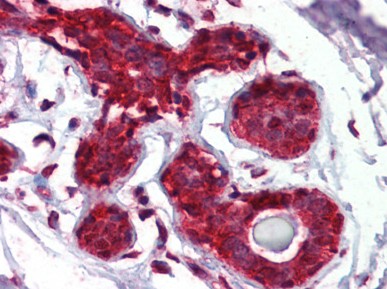
EIF4EBP1 (101-118) Rabbit Polyclonal Antibody
Other products for "EIF4EBP1"
Specifications
| Product Data | |
| Applications | EMSA, IHC, WB |
| Recommended Dilution | GS. Immunohistochemistry on Paraffin Sections: 5 - 10 µg/ml. Western Blot: 4 µg/ml. |
| Reactivities | Hamster, Human, Mouse, Rabbit, Rat |
| Host | Rabbit |
| Clonality | Polyclonal |
| Immunogen | 18 residue synthetic peptide based on the human PHAS-I (residues 101-118) and the peptide coupled to KLH |
| Specificity | This antibody reacts to 4E-binding Protein 1 (EIF4EBP1) at 101-118. |
| Formulation | BBS, pH 8.4 (25 mM sodium borate, 100 mM boric acid, 75 mM sodium chloride, 5 mM EDTA) State: Aff - Purified State: Liquid purified Ig fraction |
| Concentration | lot specific |
| Purification | Immunoaffinity Chromatography |
| Storage | Store the antibody undiluted at 2-8°C for one month or (in aliquots) at -20°C for longer. Avoid repeated freezing and thawing. |
| Stability | Shelf life: one year from despatch. |
| Gene Name | Homo sapiens eukaryotic translation initiation factor 4E binding protein 1 (EIF4EBP1) |
| Database Link | |
| Background | PHAS-I, also known as eIF4E-BP1 and PHAS-II, -III (eIF4E-BP2, 3), are members of a family of proteins that regulate eukaryotic translation initiation which is mediated by the cap structure (m7GpppN, where N=any nucleotide) present at the 5'end of all cellular mRNAs, except organellar. The m7 cap is essential for the translation of most mRNA because it directs the translation machinery of the 5' end of the mRNA via its interaction with the cap binding protein, the translation initiation factor 4E(eIF4E). eIF4E plays an principal role in determining global translation rates because its interaction with the cap facilitates the binding of the ribosome to the mRNA. Consistent with this role, eIF4E is required for cell cycle progression, exhibits anti-apoptotic activity and when overexpressed transforms cells. Interaction with PHAS proteins prevents incorporation of eIF4E into an active translation initiation complex and inhibits cap-dependent translation. However, this inhibitory effect is alleviated following phosphorylation of the PHAS proteins by a P13K-dependent pathway, involving signaling by the antiapoptotic kinase Akt/PKB, as well as FRAP/mTOR. Rat PHAS-I has 117 amino acids with a apparent molecular weight of 22 kDa and is 93% identical to eIF-4E-BPI cloned from human placenta. PHAS-I and -II were found to have overlapping but different patterns of expression in tissues. PHAS-I is expressed in a wide variety of cell types with the highest being in two of the most insulin-responsive tissues, adipocytes and skeletal muscle. Both PHAS proteins are phosphorylated in response to insulin or growth factors such as EGF, PDGF and IGF-1. Increasing cAMP in cells promotes dephosphorylation of both PHAS-I and PHAS-II but that regulation of the two protein differ because PHAS-II, unlike PHAS-I is readily phosphorylated by PKA. The PHAS-I initiation factor has 2-8 phosphorylation sites and is multiply phosphorylated by insulinstimulated protein kinase(s) resulting in 8-10 phosphorylated isoforms in exponentially growing cells. Changes occur in the expression of these isoforms in response to stresses such as heat shock, and this may contribute to translation repression. |
| Synonyms | PHAS-I, PHAS-1, PHAS1 |
| Reference Data | |
| Protein Pathways | Acute myeloid leukemia, ErbB signaling pathway, Insulin signaling pathway, mTOR signaling pathway |
Documents
| Product Manuals |
| FAQs |
{0} Product Review(s)
0 Product Review(s)
Submit review
Be the first one to submit a review
Product Citations
*Delivery time may vary from web posted schedule. Occasional delays may occur due to unforeseen
complexities in the preparation of your product. International customers may expect an additional 1-2 weeks
in shipping.


 Germany
 Japan
Japan
 United Kingdom
 China
China


