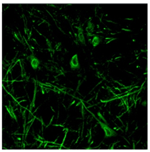
Grm5 (C-term) Rabbit Polyclonal Antibody
Other products for "Grm5"
Specifications
| Product Data | |
| Applications | IF, IHC, WB |
| Recommended Dilution | Western blot: 0.1-0.5 µg/ml. ECL on Rat microsmoal preparation. Reacts with a band of ~132 kDa. Immunocytochemistry: 0.5 µg/ml (See Protocols for more details). Immunohistochemistry: 0.1-0.2 µg/ml (See Protocols for more details). |
| Reactivities | Canine, Mouse, Rat |
| Host | Rabbit |
| Isotype | IgG |
| Clonality | Polyclonal |
| Immunogen | Synthetic peptide from C-terminus of Rat mGluR5 |
| Specificity | This antibody recognizes mGluR5 at C-term. |
| Formulation | 0.02M Phosphate, 0.2M Sodium Chloride, pH 7.6 State: Aff - Purified State: Liquid purified IgG fraction Preservative: 0.09% Sodium Azide |
| Concentration | lot specific |
| Purification | Afiinity Chromatography |
| Storage | Store the antibody undiluted (in aliquots) at -20°C Antibody may have become trapped in top of vial during shipping. Centrifugation of vial is recommended before opening. Avoid repeated freezing and thawing. |
| Stability | Shelf life: 6 months from despatch. |
| Gene Name | Rattus norvegicus glutamate metabotropic receptor 5 (Grm5) |
| Database Link | |
| Background | Metabotropic glutamate receptors (mGluRs) are G-protein coupled receptors activated by glutamate. Based on sequence similarity, transduction mechanisms and agonist potencies, mGluRs are subdivided into three groups: mGluR1/mGluR5, mGluR2/mGluR3, and mGluR4/mGluR6/mGluR7/mGluR8. mGluRs are widely distributed throughout the nervous system and are expressed by both neurons and glial cells. mGluRs have been suggested to play a variety of functional roles, among which is involvement in synaptic plasticity underlying learning and memory as well as chronic pain. |
| Synonyms | Metabotropic glutamate receptor 5, GPRC1E, MGLUR5 |
| Note | Protocol: Immunohistochemistry: Rat brain and mouse brain sections fixed with 4% paraformaldehyde containing 15% saturated picric acid in 0.1M PB, pH 7.4. Slide-mounted tissue sections were processed for indirect Immunofluorescence. Slides were incubated with blocking buffer for 1 hour at room temperature. Primary antiserum was diluted with blocking buffer to the appropriate working concentration. Blocking buffer was removed and slides were incubated for 18-24 hours at 4ºC with primary antiserum. Slides were rinsed 3 times and then incubated with secondary antibodies for 1 hour at room temperature. Slides were again rinsed 3 times and coverslipped. Staining was examined using Fluorescence microscopy. |
| Reference Data | |
Documents
| Product Manuals |
| FAQs |
{0} Product Review(s)
0 Product Review(s)
Submit review
Be the first one to submit a review
Product Citations
*Delivery time may vary from web posted schedule. Occasional delays may occur due to unforeseen
complexities in the preparation of your product. International customers may expect an additional 1-2 weeks
in shipping.


 Germany
 Japan
Japan
 United Kingdom
 China
China


