


 Islands")


")


")


")


")
")


 Germany
 Japan
Japan
 United Kingdom
 China
China
Thy1 Rabbit Polyclonal Antibody
Product Images
Other products for "Thy1"
Specifications
| Product Data | |
| Applications | Assay, CT, IHC |
| Recommended Dilution | Cytotoxicity studies (See Protocols). Immunohistochemistry on Frozen Sections: This antibody has been reported to work at 1/20 dilution. |
| Reactivities | Mouse |
| Host | Rabbit |
| Clonality | Polyclonal |
| Immunogen | CBA Brain. |
| Specificity | The antiserum is strongly cytotoxic to T-lymphocytes, and has been thoroughly adsorbed to remove cross-reactivity against B-lymphocytes. |
| Formulation | State: Serum State: Lyophilized (0.8 µm filtred, non-sterile) Serum. |
| Reconstitution Method | Restore with distilled water to initial volume. |
| Gene Name | Mus musculus thymus cell antigen 1, theta (Thy1) |
| Database Link | |
| Synonyms | Thy-1, THY1, CDw90 |
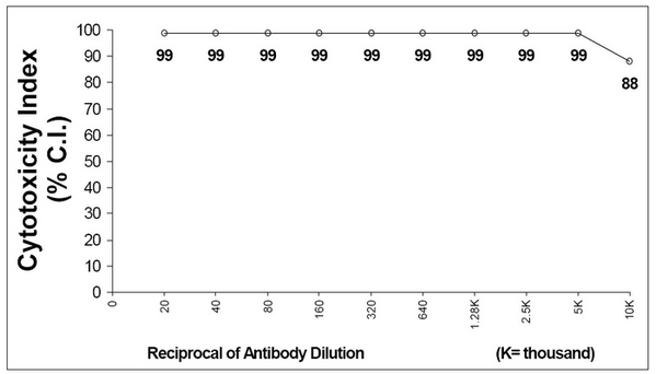
| Note | Heat Inactivation: 45 minutes at 56°C. Adsorptions: Mouse liver and peripheral blood. Protocol: Cytotoxicity Analysis: Method: 1. Prepare a cell suspension from the appropriate tissue in Cytotoxicity Mediuma or equivalent. Remove erythrocytes and dead cells (where necessary) by purification of viable lymphocytes on Lympholyte®-Mb cell separation medium. After washing, adjust the cell concentration to 1x10e6 cells per ml in Cytotoxicity Medium. 2. Add the antibody to a final concentration of 1:20 and mix. 3. Incubate for 60 minutes at 4°C. 4. Centrifuge to pellet the cells and discard the supernatant. 5. Resuspend to the original volume in Low-Tox®-M Rabbit Complementc diluted to the recommended concentration in Cytotoxicity Medium. 6. Incubate for 60 minutes at 37°C. 7. Place on ice. 8. Add Trypan Blue, 10% by volume of 1% Trypan Blue (w/v) 3-5 minutes before scoring. Score live versus dead cells in a hemacytometer. Cytotoxic Index (C. I.) can be calculated as follows: C.I. = 100%Cyt (Ab+Complement) - %Cyt (Complement) / 100% - %Cyt (Complement). Results: Antibody Titration by Cytotoxicity Analysis: Cell Source: Thymus Donor: C3H/He Cell Concentration: 1.1x10e6 cells/ml Complement: Low-Tox®-M Rabbit Complement Complement Concentration: 1:18 Procedure: Two-stage cytotoxicity as described before C.I. = 100%Cyt (Ab+Complement) - %Cyt (Complement) / 100% - %Cyt (Complement). Tissue Distribution by Cytotoxicity Analysis: Procedure: see below Antibody Concentration Used: 1:40 Strain: C3H/He Cell Source (C.I.): Thymus (100) Spleen (39) Lymph Node (75) Bone Marrow (41) Cytotoxicity Depleting Assay: Method: 1. Prepare a cell suspension from the appropriate tissue in Cytotoxicity Medium or equivalent. Remove erythrocytes and dead cells (where necessary) by purification of viable lymphocytes on Lympholyte®- M cell separation medium. After washing, adjust the cell concentration to 1x10e7 cells per ml in Cytotoxicity Medium. 2. Add the antibody to a final concentration of 1:20 and mix. Alternatively, pellet the cells and resuspend in antibody diluted 1:20 in Cytotoxicity Medium. 3. Incubate for 60 minutes at 4°C. 4. Centrifuge to pellet the cells and discard the supernatant. 5. Resuspend to the original volume in Low-Tox-M® Rabbit Complement, diluted to the appropriate concentration in Cytotoxicity Medium. (Recommended concentration included with each batch of Low-Tox-M® Rabbit Complement.) 6. Incubate for 60 minutes at 37°C. 7. Monitor for percent cytotoxicity at this stage, before further processing. For this purpose, remove a small sample from each tube, dilute 1:10 with medium, and add 1/10 volume of 1% Trypan Blue. After 3-5 minutes, score live versus dead cells in a hemacytometer. 8. For functional studies, remove the dead cells from the treated groups before further processing, particularly if the treated cells are to be cultured. Layering the cell suspension over a separation medium and centrifuging at room temperature as per the instructions provided can do this. Live cells will form a layer at the interface, while the dead cells pellet. The interface can then be collected and washed in Cytotoxicity Medium before being resuspended in the appropriate medium for further processing. Alternatively, the cells can be washed and resuspended in the appropriate medium for further processing immediately after Step #6, provided that the dead cells will not interfere with subsequent assays. Functional Testing: Method: Cells were treated as described in Cytotoxicity Depletion Assay. Treated cells and controls were tested for: a) the ability to generate plaque-forming cells (PFC) using a modified Jerne haemolytic plaque assay. b) the ability to generate cytotoxic T effector cells using a cytotoxic lymphocyte reaction (CTL) assay. Cells were treated both before and after sensitization in the CTL assay. Results: Cell Source: Splenocytes Donors: C3H/He and BALB/c Cell Concentration: 1x10e7 cells/ml. Antibody Concentration Used: 1:40 Complement: Low-Tox®-M Rabbit Complement Complement Concentration Used: 1:20 Treatment of C3H/He and BALB/c splenocytes with AP31644SU plus complement resulted in a significant reduction in the number of plaque-forming cells. Cytotoxic T cell function was essentially eliminated in both pre-sensitized and post-sensitized treated samples. These results are consistent with the removal of T helper and T cytotoxic cell activity. Mitogen Response: Method: Cells were treated as described in Cytotoxicity Depletion Assay. Remaining viable lymphocytes were exposed to the mitogens Concanavalin A (CON A), Phytohaemagglutinin (PHA) and Lipopolysaccharide (LPS). Results: Cell Source: C3H/He splenocytes Cell Concentration: 1.1x10e7 cells/ml Antibody Concentration Used: 1:80 Complement: Low-Tox®-M Rabbit Complement Complement Concentration: 1:20 Cell depletion with AP31644SU had little effect on the LPS response (< 5%) and essentially eliminated the CON A and PHA response (98% and 96%, respectively). Notes: a. Cytotoxicity Medium is RPMI-1640 with 25 mM Hepes buffer and 0.3% bovine serum albumin (BSA). BSA is substituted for the conventionally used fetal calf serum (FCS) because we have found that many batches of FCS contain complement-dependent cytotoxins to mouse lymphocytes, thus increasing the background killing in the presence of complement. Some batches of BSA also contain complement-dependent cytotoxins, resulting in the same problem. We screen for batches of BSA giving low background in the presence of complement and use the selected BSA for preparing Cytotoxicity Medium. b. Lympholyte®-M cell separation medium is a density separation medium designed specifically for the isolation of viable mouse lymphocytes. This separation medium provides a high and non-selective recovery of viable mouse lymphocytes, removing erythrocytes and dead cells. The density of this medium is 1.087-1.089. Isolation of mouse lymphocytes on cell separation medium of density 1.077 will result in high and selective loss of lymphocytes and should be avoided. c. Rabbit serum provides the most potent source of complement for use with antibodies to mouse cell surface antigens. However, rabbit serum itself is very toxic to mouse lymphocytes. Low-Tox®-M Rabbit Complement is absorbed to remove toxicity to mouse lymphocytes, while maintaining its high complement activity. When used in conjunction with Cytotoxicity Medium, this reagent provides a highly potent source of complement with minimal background toxicity. |
| Reference Data | |
Documents
| Product Manuals |
| FAQs |
| SDS |
{0} Product Review(s)
0 Product Review(s)
Submit review
Be the first one to submit a review


Product Citations
*Delivery time may vary from web posted schedule. Occasional delays may occur due to unforeseen
complexities in the preparation of your product. International customers may expect an additional 1-2 weeks
in shipping.