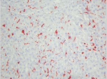
Adgre1 Rat Monoclonal Antibody [Clone ID: BM8]
Other products for "Adgre1"
Specifications
| Product Data | |
| Clone Name | BM8 |
| Applications | FC, IF, IHC |
| Recommended Dilution | Immunohistochemistry on Frozen Sections: 0.4 μg/ml (1/1000). Immunohistochemistry on Paraffin Sections: 4 μg/ml (1/100). Proteinase K pretreatment for antigen retrieval is recommended. Positive Control: Mouse spleen. Has been described to work in FACS. |
| Reactivities | Mouse |
| Host | Rat |
| Isotype | IgG2a |
| Clonality | Monoclonal |
| Immunogen | Cultured macrophages. The F4/80 antigen is a 125 kD extracellular membrane protein sensitive to 2-Mercaptoethanol. |
| Specificity | This antibody is useful for the detection of major subpopulations of resident tissue macrophages. The antigen expression increases upon maturation of macrophage precursors in bone marrow and blood as well as in ontogeny. This clone BM8 is the only macrophage marker that is able to distinguish non-destructive from destructive inflammation processes in the pancreas and has been shown to be a unique histological marker of the progression from peri-insulitis to ß-cell destruction and diabetes in a mouse diabetes model. Antigen Distribution on Tissue and Isolated Sections: The antigen is detected on tissue fixed macrophages in all organs tested so far (spleen, lymph nodes, thymus, liver, skin). It is also present on Langerhans cells in the skin and Kupffer cells in the liver. In complete Freund's adjuvant induced granulomas the antigen is expressed by inflammatory macrophages, but is absent from epitheloid cells. The antigen is expressed in vitro on over 80% of M-CSF stimulated bone marrow derived macrophages, after a few days of culture. It is absent from granulocytes, lymphocytes and thrombocytes. |
| Formulation | Phosphate buffered saline pH 7.2 (PBS) State: Purified State: Lyophilized purified IgG fraction Stabilizer: 5 mg/ml BSA Preservative: 0.05% (v/v) Kathon CG |
| Reconstitution Method | Restore by adding 0.5 ml distilled water |
| Concentration | 0.4 mg/ml (after reconstitution) |
| Purification | Immunoaffinity Chromatography |
| Storage | Store lyophilized at 2-8°C for 6 months or at -20°C long term. After reconstitution store the antibody undiluted at 2-8°C for one month or (in aliquots) at -20°C long term. Avoid repeated freezing and thawing. |
| Stability | Shelf life: one year from despatch. |
| Gene Name | Mus musculus adhesion G protein-coupled receptor E1 (Adgre1) |
| Database Link | |
| Background | F4/80 antigen is a 160 kDa cell surface glycoprotein that is a member of the EGF TM7 family of proteins which shares 68% overall amino acid identity with human EGF module containing mucin like hormone receptor 1 (EMR1). Expression of F4/80 is heterogeneous and is reported to vary during macrophage maturation and activation. The F4/80 antigen is expressed on a wide range of mature tissue macrophages including Kupffer cells, Langerhans, microglia, macrophages located in the gut lamina propria, peritoneal cavity, lung, thymus, bone marrow stroma and macrophages in the red pulp of the spleen. F4/80 expression has also been reported on a subpopulation of dendritic cells but is absent from macrophages located in T cell areas of the spleen and lymph node. The ligands and biological functions of the F4/80 antigen have not yet been determined but recent studies suggest a role for F4/80 in the generation of efferent CD8+ve regulatory T cells. |
| Synonyms | Emr1, Gpf480 |
| Note | Protocol: Protocol with frozen, ice-cold acetone-fixed sections: The whole procedure is performed at room temperature 1. Wash in PBS 2. Block endogenous peroxidase 3. Wash in PBS 4. Block with 10% normal goat serum in PBS for 30min. in a humid chamber 5. Incubate with primary antibody (dilution see datasheet) for 1h in a humid chamber 6. Wash in PBS 7. Incubate with secondary antibody (peroxidase-conjugated goat anti rat IgG (H+L) minimal-cross reaction to mouse) for 1h in a humid chamber 8. Wash in PBS 9. Incubate with AEC substrate (3-amino-9-ethylcarbazol) for 12min. 10. Wash in PBS 11. Counterstain with Mayer’s hemalum. Protocol with formalin-fixed, paraffin-embedded sections: The whole procedure is performed at room temperature 1. Deparaffinize and rehydrate tissue section 2. Incubate the tissue section with proteinase K for 7min. 3. Wash in distilled water 4. Block endogenous peroxidase 5. Wash in PBS 6. Block with 10% normal goat serum in PBS for 30min. in a humid chamber 7. Incubate with primary antibody (dilution see datasheet) for 1h in a humid chamber 8. Wash in PBS 9. Incubate with secondary antibody (peroxidase-conjugated goat anti rat IgG (H+L) minimal-cross reaction to mouse) for 1h in a humid chamber 10. Wash in PBS 11. Incubate with AEC substrate (3-amino-9-ethylcarbazol) for 12min. 12. Wash in PBS 13. Counterstain with Mayer’s hemalum. |
| Reference Data | |
Documents
| Product Manuals |
| FAQs |
{0} Product Review(s)
0 Product Review(s)
Submit review
Be the first one to submit a review
Product Citations
*Delivery time may vary from web posted schedule. Occasional delays may occur due to unforeseen
complexities in the preparation of your product. International customers may expect an additional 1-2 weeks
in shipping.


 Germany
 Japan
Japan
 United Kingdom
 China
China


