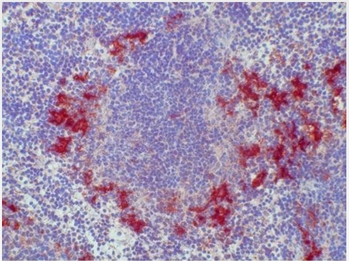
Cd209b Rat Monoclonal Antibody [Clone ID: ER-TR9]
Specifications
| Product Data | |
| Clone Name | ER-TR9 |
| Applications | FC, IHC |
| Recommended Dilution | Immunohistochemistry on Frozen Sections: 1.5 µg/ml (1/200). Suggested Positive Control: Mouse spleen. Does not react on routinely processed Paraffin Sections. Has been reported to work in Flow Cytometry (Use 10 µl stock solution to label 106 cells). |
| Reactivities | Mouse |
| Host | Rat |
| Isotype | IgM |
| Clonality | Monoclonal |
| Immunogen | Thymus cells. |
| Specificity | Antigen, Epitope: The antigen is a Glutaraldehyde (0.05%) resistant protein expressed in the cytoplasm and on the cell surface. Monoclonal antibody ER-TR9 is a very useful marker for the identification of macrophage subpopulations present in the marginal zone of spleen and lymph node medulla. In combination with MOMA-1 (Cat#SM066P), the murine metallophilic macrophage marker, a detailed characterization of murine splenic marginal zone macrophages is obtained. ER-TR9 is also useful when studying phagocytosis of polysaccharides since the antibody selectively inhibits uptake of these glycans by macrophages. The antigen recognized by ER-TR9 has recently been shown to be the murine analogue of the human DC-SIGN (Dendritic Cell - Specific ICAM-3 Grabbing Non-Integrin), named SIGN-R1. The antibody reacts with a Mouse subpopulation of mature tissue macrophages present in the splenic marginal zone, lymph node medullary and trabecular sinuses. Antigen Distribution Isolated Cells: The antigen is found on a subpopulation of phagocytic macrophages isolated from the spleen and showing acid phosphatase and moderate non-specific esterase activity. These phagocytes selectively ingest neutral polysaccharides such as Ficoll. Tissue Sections: Subpopulation of resident macrophages in the splenic marginal zone which are in the proximity of a certain B cell subpopulation (µ+, d-). It is also found on a subpopulation of macrophages localized in the medullary sinuses and trabecular sinuses of lymph nodes. Furthermore, macrophage subpopulations in other organs, such as some connective tissue macrophages in the dermis, may also show ER-TR9 antigen expression. |
| Formulation | Stock solution contains PBS, pH 7.2 with 5 mg/ml BSA as stabilizer and 0.09% Sodium Azide as a preservative Label: Biotin State: Lyophilized purified Ig fraction |
| Reconstitution Method | Restore with 0.5 ml distilled water (= 0.3 mg/ml Stock Solution). |
| Concentration | 0.3 mg/ml (after reconstitution) |
| Purification | Affinity Chromatography |
| Conjugation | Biotin |
| Storage | Store the original vial at 2-8°C up to one year. Stock solution or aliquots thereof can be stored for 6 months at 2-8°C. Do not freeze working dilutions. |
| Database Link | |
| Background | SIGN R1 is a specific marker for the identification of macrophage subpopulations present in the marginal zone of spleen (the so-called marginal zone macrophages (MZM)), in the lymph node medulla, and in some strains, in the peritoneal cavity. MZM of the spleen are involved in the clearance of polysaccharides. Mouse SIGN R1 is a C type lectin, like DC SIGN which is expressed on Human dendritic cells (DCs). However, Mmouse SIGN R1 itself is not expressed on DCs. SIGN R1 exists in an aggregated form, resistant to dissociation into monomers upon boiling in SDS under reducing conditions. SIGN R1 mediates the uptake of encapsulated organisms and may be an important mediator for the uptake of microbes in both spleen and lymph node, particularly through the recognition of microbial polysaccharides. |
| Synonyms | DC-SIGN-related protein 1, OtB7 |
| Reference Data | |
Documents
| Product Manuals |
| FAQs |
{0} Product Review(s)
0 Product Review(s)
Submit review
Be the first one to submit a review
Product Citations
*Delivery time may vary from web posted schedule. Occasional delays may occur due to unforeseen
complexities in the preparation of your product. International customers may expect an additional 1-2 weeks
in shipping.


 Germany
 Japan
Japan
 United Kingdom
 China
China


