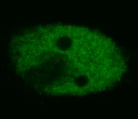
p53 (TP53) (16-25) Mouse Monoclonal Antibody [Clone ID: BP53-12]
Specifications
| Product Data | |
| Clone Name | BP53-12 |
| Applications | ELISA, IF, IHC, IP, WB |
| Recommended Dilution | ELISA. Western Blotting (Non-reducing conditions): Recommended Dilution: 1-2 µg/ml, overnight in 4°C. Positive Control: RAMOS Human lymphoma cell line Sample Preparation: Resuspend approx. 50 mil. cells in 1 ml cold Lysis buffer (1% laurylmaltoside in 20 mM Tris/Cl, 100 mM NaCl pH 8.2, 50 mM NaF including Protease inhibitor Cocktail). Incubate 60 min on ice. Centrifuge to remove cell debris. Mix lysate with non-reducing SDS-PAGE sample buffer. SDS-PAGE: 12% separating gel. Immunoprecipitation. Immunocytochemistry. Immunohistochemistry on Frozen and Paraffin-Embedded Sections. |
| Reactivities | Human, Primate |
| Host | Mouse |
| Isotype | IgG2a |
| Clonality | Monoclonal |
| Immunogen | Bacterially expressed full-length wild-type p53. |
| Specificity | This Monoclonal antibody BP53-12 recognizes defined epitope (aa 16-25) on Human p53, a 50 kDa tumour suppressor found in increased amounts in a wide variety of transformed cells. It is frequently mutated or inactivated in many types of cancer. |
| Formulation | Phosphate buffered saline (PBS), pH~7.4 State: Purified State: Liquid purified Ig fraction (> 95% pure by SDS-PAGE) Preservative: 15 mM Sodium Azide |
| Concentration | lot specific |
| Purification | Precipitation Methods |
| Storage | Store undiluted at 2-8°C for one month or (in aliquots) at -20°C for longer. Avoid repeated freezing and thawing. |
| Stability | Shelf life: one year from despatch. |
| Database Link | |
| Background | The tumour suppressor protein p53 is a key element of intracellular anticancer protection. It mediates cell cycle arrest or apoptosis in response to DNA damage or to starvation for pyrimidine nukleotides. It is up-regulated in response to these stress signals and stimulated to activate transcription of specific genes, resulting in expression of p21waf1 and other proteins involved in G1 or G2/M arrest, or proteins that trigger apoptosis, such as Bcl-2. The structure of p53 comprises N-terminal transactivation domain, central DNA-binding domain, oligomerisation domain, and C-terminal regulatory domain. There are various phosphorylation sites on p53, of which the phosphorylation at Ser15 is important for p53 activation and stabilization. |
| Synonyms | Cellular tumor antigen p53, Tumor suppressor p53, Phosphoprotein p53, NY-CO-13 |
| Reference Data | |
Documents
| Product Manuals |
| FAQs |
{0} Product Review(s)
0 Product Review(s)
Submit review
Be the first one to submit a review
Product Citations
*Delivery time may vary from web posted schedule. Occasional delays may occur due to unforeseen
complexities in the preparation of your product. International customers may expect an additional 1-2 weeks
in shipping.


 Germany
 Japan
Japan
 United Kingdom
 China
China


