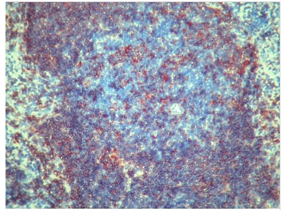
MHC Class II (I-A k,b,d,q,r) Rat Monoclonal Antibody [Clone ID: ER-TR3]
Specifications
| Product Data | |
| Clone Name | ER-TR3 |
| Applications | FC, IHC |
| Recommended Dilution | Immunohistochemistry on Frozen Sections: 1.5 µg/ml (1/200). Suggested Positive Control: Mouse spleen. Does not react on routinely processed paraffin sections. Has been reported to work in Flow Cytometry. |
| Reactivities | Mouse |
| Host | Rat |
| Isotype | IgG2b |
| Clonality | Monoclonal |
| Immunogen | Murine thymic reticulum. |
| Specificity | Monoclonal antibody ER-TR3 detects MHC class II antigens encoded by the murine Ia region of the H-2 complex, corresponding to the human HLA-DR region. it is a valuable tool for studying T-helper cell interaction with class II positive antigen presenting cells (dendritic cells, B-cells, macrophages). This antibody also offers new possibilities for studying the development of T-helper cells since it also stains stromal cells in the thymus. Antigen Distribution Isolated Cells: The antigen is found on dendritic cells, B-cells and macrophages. Tissue sections: The antigen is found on B-cells, interdigitating cells and macrophages in peripheral lymphoid organs but is absent from T-cells. It is also expressed as a fine reticular pattern on stromal thymic cells of the cortex and as a confluent pattern on stromal thymic cells of the medulla. |
| Formulation | Stock Solution contains PBS, pH 7.2 with 5 mg/ml BSA as a stabilizer and 0.09% Sodium Azide as preservative State: Purified State: Lyophilized purified Ig fraction |
| Reconstitution Method | Restore with 0.5 ml distilled water. |
| Concentration | 0.3 mg/ml |
| Purification | Affinity Chromatography |
| Storage | Store lyophilized at 2-8°C and reconstituted at -20°C. Avoid repeated freezing and thawing. |
| Stability | Shelf life: One year from despatch. |
| Background | MHC Class II antigens are heterodimers consisting of one alpha chain (31-34kD) and one beta chain (26-29kD). The family of monoclonal antibodies (ER-TR3, ER-TR2, ER-TR1) detect MHC class II antigens encoded by the murine Ia region of the H-2 complex, corresponding to the Human HLA-DR region. MHC Class II antigens are a valuable tool for studying T helper cell interaction with class II positive antigen presenting cells (dendritic cells, B cells, macrophages) and offer new possibilities for studying the development of T helper cells since these antibodies also stain stromal cells in the thymus. MHC Class II antigens are also inducible on a number of other cells (endothelium and epithelial cells) by interferon gamma. |
| Note | Protocol: Protocol with Frozen, ice-cold Acetone-Fixed Sections: The whole procedure is performed at room temperature 1. Wash in PBS 2. Block endogenous peroxidase 3. Wash in PBS 4. Block with 10 % normal goat serum in PBS for 30 min. in a humid chamber 5. Incubate with primary antibody (dilution see datasheet) for 1h in a humid chamber 6. Wash in PBS 7. Incubate with secondary antibody (peroxidase-conjugated goat anti rat IgG (H+L) minimal-cross reaction to mouse) for 1h in a humid chamber 8. Wash in PBS 9. Incubate with AEC substrate (3-amino-9-ethylcarbazol) for 12 min. 10. Wash in PBS 11. Counterstain with Mayer's hemalum |
| Reference Data | |
Documents
| Product Manuals |
| FAQs |
{0} Product Review(s)
0 Product Review(s)
Submit review
Be the first one to submit a review
Product Citations
*Delivery time may vary from web posted schedule. Occasional delays may occur due to unforeseen
complexities in the preparation of your product. International customers may expect an additional 1-2 weeks
in shipping.


 Germany
 Japan
Japan
 United Kingdom
 China
China


