


 Islands")


")


")


")


")
")


 Germany
 Japan
Japan
 United Kingdom
 China
China
Cd8a Mouse Monoclonal Antibody [Clone ID: 49-31.1]
Product Images
 can be calculated as follows:")


Specifications
| Product Data | |
| Clone Name | 49-31.1 |
| Applications | Assay, CT, FC |
| Recommended Dilution | Flow Cytometry Analysis. Cytotoxicity Analysis (See Protocols). Functional Testing (See Protocols). Cytotoxicity Depletion Assays (See Protocols). |
| Reactivities | Mouse |
| Host | Mouse |
| Isotype | IgG3 |
| Clonality | Monoclonal |
| Immunogen | Recipient: 129/ReJ. Donor: CBA. Fusion Partner: Spleen from immunized recipient fused with Myeloma P3 NSI-Ag 4-1. |
| Specificity | This Ly-2.1 Monoclonal Antibody reacts with a sub-population of lymphocytes from mouse strains expressing the Ly 2.1 (CD8a) phenotype but does not react with lymphocytes from mouse strains expressing the Ly 2.2 phenotype. |
| Formulation | State: Ascites State: Lyophilized Ascites |
| Reconstitution Method | Restore with 0.5 ml of distilled water. |
| Database Link | |
| Background | The CD8 antigen is a cell surface glycoprotein found on most cytotoxic T lymphocytes that mediates efficient cell to cell interactions within the immune system. The CD8 antigen, acting as a coreceptor, and the T cell receptor on the T lymphocyte recognize antigen displayed by an antigen presenting cell (APC) in the context of class I MHC molecules. The functional coreceptor is either a homodimer composed of two alpha chains, or a heterodimer composed of one alpha and one beta chain. Both alpha and beta chains share significant homology to immunoglobulin variable light chains. |
| Synonyms | CD8 alpha chain, CD8A, MAL |
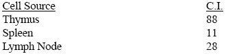
| Note | Sterility: This reagent is not sold as sterile, but can be sterilized by filtration if necessary. To minimize loss of volume during filtration, dilute to the final working concentration in the appropriate medium before filtration. Protocol: CYTOTOXICITY ANALYSIS: Method for Determining percent of Ly 2.1+ cells in a population: 1. Prepare a cell suspension from the appropriate tissue in Cytotoxicity Medium or equivalent. Remove red cells and dead cells (where necessary) by purification of viable lymphocytes on Lympholyte®-M density cell separation medium. After washing, adjust the cell concentration to 1x10e6 cells per ml in Cytotoxicity Medium. 2. Add the antibody to a final concentration of 1:1000 and mix. 3. Incubate for 60 minutes at 4°C. 4. Centrifuge to pellet the cells and discard the supernatant. 5. Resuspend to the original volume in Low-Tox®-M Rabbit Complement diluted to the recommended concentration in Cytotoxicity Medium. 6. Incubate for 60 minutes at 37°C. 7. Place on ice. 8. Add Trypan Blue, 10% by volume of 1% Trypan Blue (w/v) added 3-5 minutes before scoring works well. Score live versus dead cells in a hemacytometer. Results - Antibody Titration: Cell Source: Thymus Donor: B6-Ly-2a3a (Ly 2.1) Cell Concentration: 1x10e6 cells/ml Complement: Low-Tox®-M Rabbit Complement Complement Concentration: 1:18 Procedure: Two-stage cytotoxicity Results - Tissue Distribution: Antibody Concentration Used: 1:500 Strain: B6-Ly-2a3a Results - Strain Distribution: Antibody Concentration Used: 1:500 Strains Tested: C57BL/6, C3H/He, B6-Ly-2a3a, CBA/J, BALB/c,DBA/2, A.TH Positive: B6-Ly-2a3a , CBA/J, DBA/2, C3H/He Negative: C57BL/6, BALB/c, A.TH CYTOTOXICITY DEPLETION ASSAY: Method for depleting a Ly 2.1+ cell population for further functional analysis: 1. Prepare a cell suspension from the appropriate tissue in Cytotoxicity Mediuma or equivalent. Remove red cells and dead cells (where necessary) by purification of viable lymphocytes on Lympholyte®-M density cell separation medium . After washing, adjust the cell concentration to 1x10e6 cells per ml in Cytotoxicity Medium. 2. Add the antibody to a final concentration of 1:500 and mix. Alternatively, pellet the cells and resuspend in antibody diluted 1:500 in Cytotoxicity Medium. 3. Incubate for 60 minutes at 4°C. 4. Centrifuge to pellet the cells and discard the supernatant. 5. Resuspend to the original volume in Low-Tox-M® Rabbit Complementc, diluted to the appropriate concentration in Cytotoxicity Medium. (Recommended concentration included with each batch of Low-Tox-M® Rabbit Complement.) 6. Incubate for 60 minutes at 37°C. 7. Monitor for percent cytotoxicity at this stage, before further processing. For this purpose, remove a small sample from each tube, dilute 1:10 with medium, and add 1/10 volume of 1% Trypan Blue. After 3-5 minutes, score live versus dead cells in a hemacytometer. 8. For functional studies, remove the dead cells from the treated groups before further processing, particularly if the treated cells are to be cultured. This can be done by layering the cell suspension over a separation medium and centrifuging at room temperature as per the instructions provided. Live cells will form a layer at the interface, while the dead cells pellet. The interface can then be collected and washed in Cytotoxicity Medium before being resuspended in the appropriate medium for further processing. Alternatively, the cells can be washed and resuspended in the appropriate medium for further processing immediately after Step #6, provided that the dead cells will not interfere with subsequent assays. FUNCTIONAL TESTING: Method: Cells were treated as described in Cytotoxicity Depletion Assay. Treated cells and controls were tested for: a) the ability to generate plaque-forming cells (PFC) using a modified Jerne haemolytic plaque assay. b) the ability to generate cytotoxic T effector cells using a cytotoxic lymphocyte reaction (CTL) assay. Cells were treated both before and after sensitization in the CTL assay. Results: Cell Source: Splenocytes Donors: C57/BL6 and C3H/He Cell Concentration: 1x10e7 cells/ml Antibody Concentration Used: 1:20 Complement: Low-Tox®-M Rabbit Complement Complement Concentration Used: 1:10 Treatment of C3H/He splenocytes with CL8921A plus complement had no effect on the number of plaque-forming cells. However, marked inhibition of cytotoxic T cell function was found to occur when cells were treated before or after sensitization in the CTL assay. This antibody blocks T cell function in the absence of complement as determined by 51Cr release T-cytotoxicity assay. No effect in either assay was observed when C57BL/6 cells were used. These results are consistent with the depletion of cytotoxic T cells of the Ly 2.1 phenotype. |
| Reference Data | |
Documents
| Product Manuals |
| FAQs |
| SDS |
{0} Product Review(s)
0 Product Review(s)
Submit review
Be the first one to submit a review


Product Citations
*Delivery time may vary from web posted schedule. Occasional delays may occur due to unforeseen
complexities in the preparation of your product. International customers may expect an additional 1-2 weeks
in shipping.