


 Islands")


")


")


")


")
")


 Germany
 Japan
Japan
 United Kingdom
 China
China
Cd44 Rat Monoclonal Antibody [Clone ID: IM7.8.1]
Product Images
Specifications
| Product Data | |
| Clone Name | IM7.8.1 |
| Applications | FC |
| Recommended Dilution | Flow Cytometry (see Protocols). (Reported to be useful in immunoprecipitation, ELISA, cytotoxicity assays and immunohistochemistry of frozen sections and complement depletion.) |
| Reactivities | Mouse |
| Host | Rat |
| Isotype | IgG2b |
| Clonality | Monoclonal |
| Specificity | Unconjugated antibody is cited in: Keittisak Suwan, Kanyamas Choocheep, Sonoko Hatano, Prachya Kongtawelert, Koji Kimata, and Hideto Watanabe. Versican/PG-M assembles hyaluronan into extracellular matrix and inhibits CD44-mediated signaling toward premature senescence in embryonic fibroblasts. J. Biol. Chem., Jan 2009; 10.1074/jbc.M806927200. |
| Formulation | PBS containing 0.09% Sodium Azide as preservative and EIA grade BSA as a stabilizing protein to bring total protein concentration to 4-5 mg/ml. Label: Biotin State: Liquid purified Ig fraction |
| Concentration | 0.1 mg/ml |
| Conjugation | Biotin |
| Database Link | |
| Background | CD44 is a type 1 transmembrane glycoprotein also known as Phagocytic Glycoprotein 1 (pgp 1) and HCAM. CD44 is the receptor for hyaluronate and exists as a large number of different isoforms due to alternative RNA splicing. The major isoform expressed on lymphocytes, myeloid cells, and erythrocytes is a glycosylated type 1 transmembrane protein. Other isoforms contain glycosaminoglycans and are expressed on hematopoietic and non hematopoietic cells. CD44 is involved in adhesion of leukocytes to endothelial cells, stromal cells, and the extracellular matrix. |
| Synonyms | LHR, MDU2, MDU3, MIC4, CDw44, Epican, ECMR-III, HUTCH-I, Heparan sulfate proteoglycan, Hermes antigen, Hyaluronate receptor, PGP-1 |
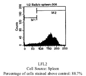
| Note | Protocol: FLOW CYTOMETRY ANALYSIS: Method: 1. Prepare a cell suspension in media A. For cell preparations, deplete the red blood cell population with Lympholyte®-M cell separation medium. 2. Wash 2 times. 3. Resuspend the cells to a concentration of 2x10e7 cells/ml in media A. Add 50 µl of this suspension to each tube (each tube will then contain 1 x 10e6 cells, representing 1 test). 4. To each tube, add ~0.25µg* of this Ab per 10e6 cells. 5. Vortex the tubes to ensure thorough mixing of antibody and cells. 6. Incubate the tubes for 30 minutes at 4°C. 7. Wash 2 times at 4°C. 8. Add 100 µl of secondary antibody (Streptavidin-PE) at a 1:50 dilution. 9. Incubate tubes at 4°C for 30 - 60 minutes (It is recommended that tubes are protected from light since most fluorochromes are light sensitive). 10. Wash 2 times at 4°C. 11. Resuspend the cell pellet in 50 µl ice cold media B. 12. Transfer to suitable tubes for flow cytometric analysis containing 15 µl of propidium iodide at 0.5 mg/ml in PBS. This stains dead cells by intercalating in DNA. Media: A. Phosphate buffered saline (pH 7.2) + 5% normal serum of host species + sodium azide (100 µl of 2M sodium azide in 100 mls). B. Phosphate buffered saline (pH 7.2) + 0.5% Bovine serum albumin + sodium azide (100 µl of 2M sodium azide in 100 mls). Results - Tissue Distribution: Mouse Strain: BALB/c Cell Concentration: 1x10e6 cells per test Antibody Concentration Used: 0.25 µg/10e6 cells Isotypic Control: Biotin Rat IgG2b |
| Reference Data | |
Documents
| Product Manuals |
| FAQs |
| SDS |
{0} Product Review(s)
0 Product Review(s)
Submit review
Be the first one to submit a review


Product Citations
*Delivery time may vary from web posted schedule. Occasional delays may occur due to unforeseen
complexities in the preparation of your product. International customers may expect an additional 1-2 weeks
in shipping.