


 Islands")


")


")


")


")
")


 Germany
 Japan
Japan
 United Kingdom
 China
China
Kit Rat Monoclonal Antibody [Clone ID: ACK4]
Product Images
Other products for "Kit"
Specifications
| Product Data | |
| Clone Name | ACK4 |
| Applications | FC |
| Recommended Dilution | Flow Cytometry. |
| Reactivities | Mouse |
| Host | Rat |
| Isotype | IgG2a |
| Clonality | Monoclonal |
| Immunogen | IL-3 dependent mast cells derived from WB- +/+ mice Donor: Wistar spleen Fusion Partner: X63.653. Ag8 |
| Specificity | Anti-mouse CD117 monoclonal antibody recognizes the receptor tyrosine kinase, c-kit. The ligand for this receptor is steel factor (stem cell factor), which exists in both soluble and membrane form. The interaction between steel factor and c-kit is essential for the development of hematopoietic, gonadal and pigment stem cells. c-kit positive cells are a subset of CD34+ hematopoietic precursor cells and it is expressed on 5-10% of total adult bone marrow cells. |
| Formulation | PBS containing 0.02% sodium azide (NaN3) as preservative and EIA grade BSA as a stabilizing protein to bring total protein concentration to 4-5 mg/ml. Label: Biotin State: Liquid purified Ig fraction |
| Concentration | 0,1 mg/ml |
| Purification | Affinity chromatography on Protein G |
| Conjugation | Biotin |
| Gene Name | Mus musculus KIT proto-oncogene receptor tyrosine kinase (Kit), transcript variant 2 |
| Database Link | |
| Synonyms | SCFR, KIT |
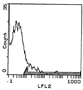
| Note | Protocol: FLOW CYTOMETRY ANALYSIS: Method: 1. Prepare a cell suspension in media A. For cell preparations, deplete the red blood cell population with Lympholyte®-M (cell separation medium). 2. Wash 2 times. 3. Resuspend the cells to a concentration of 2x10e7 cells/ml in media A. Add 50 μl of this suspension to each tube (each tube will then contain 1 x 10e6 cells, representing 1 test). 4. To each tube, add 0.2-0.1 mg of this antibody per 10e6 cells. 5. Vortex the tubes to ensure thorough mixing of antibody and cells. 6. Incubate the tubes for 30 minutes at 4°C. 7. Wash 2 times at 4°C. 8. Add 100 μl of secondary antibody (Streptavidin-PE) at a 1/250 dilution. 9. Incubate tubes at 4°C for 30 - 60 minutes (It is recommended that tubes are protected from light since most fluorochromes are light sensitive). 10. Wash 2 times at 4°C. 11. Resuspend the cell pellet in 50 μl ice cold media B. 12. Transfer to suitable tubes for flow cytometric analysis containing 15 μl of propidium iodide at 0.5 mg/ml in PBS. This stains dead cells by intercalating in DNA. Media: A. Phosphate buffered saline (pH 7.2) + 5% normal serum of host species + sodium azide (100 μl of 2M sodium azide in 100 mls). B. Phosphate buffered saline (pH 7.2) + 0.5% Bovine serum albumin + sodium azide (100 μl of 2M sodium azide in 100 mls). Results: Tissue Distribution by Flow Cytometry Analysis: Mouse Strain: C3H/He Cell Concentration: 1x10e6 cells per test Antibody Concentration Used: 0.2 μg/10e6 cells Isotypic Control: Biotin Rat IgG2a Strain Distribution by Flow Cytometry Analysis: Cell Concentration: 1x10e6 cells per test Antibody Concentration Used: 0.2 mg /10e6 cells Strains Tested: AKR, BALB/c, C3H/He, Positive: AKR, BALB/c, C3H/He, Negative: none |
| Reference Data | |
Documents
| Product Manuals |
| FAQs |
| SDS |
{0} Product Review(s)
0 Product Review(s)
Submit review
Be the first one to submit a review


Product Citations
*Delivery time may vary from web posted schedule. Occasional delays may occur due to unforeseen
complexities in the preparation of your product. International customers may expect an additional 1-2 weeks
in shipping.