Kit Rat Monoclonal Antibody [Clone ID: ACK4]
Other products for "Kit"
Specifications
| Product Data | |
| Clone Name | ACK4 |
| Applications | FC, IF, IHC |
| Recommended Dilution | Flow Cytometry: Use at 0.2 μg/ 106 cells. Reported to be useful in immunohistochemistry on Frozen and Polyester Wax Sections. |
| Reactivities | Mouse |
| Host | Rat |
| Isotype | IgG2a |
| Clonality | Monoclonal |
| Immunogen | IL-3 dependent mast cells derived from WB- +/+ mice. Donor: Wistar spleen. Fusion Partner: X63.653. Ag8. |
| Specificity | This monoclonal antibody recognizes the receptor tyrosine kinase, c-kit. The ligand for this receptor is steel factor (stem cell factor), which exists in both soluble and membrane form. The interaction between steel factor and c-kit is essential for the development of hematopoietic, gonadal and pigment stem cells. c-kit positive cells are a subset of CD34+ hematopoietic precursor cells and it is expressed on 5-10% of total adult bone marrow cells. |
| Formulation | PBS containing 0.02% Sodium Azide as preservative. State: Purified State: Liquid purified IgG fraction. |
| Concentration | 1.0 mg/ml |
| Purification | Protein G Chromatography. |
| Gene Name | Mus musculus KIT proto-oncogene receptor tyrosine kinase (Kit), transcript variant 2 |
| Database Link | |
| Background | c-Kit is a transmembrane tyrosine kinase encoded by the cKit proto oncogene. c-Kit acts to regulate a variety of biological responses including cell proliferation, apoptosis, chemotaxis and adhesion. Ligand binding to the extracellular domain leads to autophosphorylation on several tyrosine residues within the cytoplasmic domain, and activation. Mutations in c-Kit have been found to be important for tumor growth and progression in a variety of cancers including mast cell diseases, gastrointestinal stromal tumor, acute myeloid leukemia, Ewing sarcoma and lung cancer. Phosphorylation at tyrosine 721 of c-Kit allows binding and activation of PI3 kinase. |
| Synonyms | SCFR, KIT |
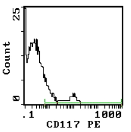
| Note | Protocol: FLOW CYTOMETRY ANALYSIS: Method: 1. Prepare a cell suspension in media A. For cell preparations, deplete the red blood cell population with Lympholyte®-M cell separation medium. 2. Wash 2 times. 3. Resuspend the cells to a concentration of 2x10e7 cells/ml in media A. Add 50 µl of this suspension to each tube (each tube will then contain 1x10e6 cells, representing 1 test). 4. To each tube, add 0.2-0.1 µg of CL042P or CL042PX per 10e6 cells. 5. Vortex the tubes to ensure thorough mixing of antibody and cells. 6. Incubate the tubes for 30 minutes at 4°C. 7. Wash 2 times at 4°C. 8. Add 100 µl of PE Goat anti-Rat IgG (H+L) secondary antibody at appropriate dilution. 9. Incubate the tubes at 4°C for 30-60 minutes. (It is recommended that the tubes are protected from light since most fluorochromes are light sensitive). 10. Wash 2 times at 4°C in media B. 11. Resuspend the cell pellet in 50 µl ice cold media B. 12. Transfer to suitable tubes for flow cytometric analysis containing 15 µl of propidium iodide at 0.5 mg/ml in PBS. This stains dead cells by intercalating in DNA. Media: A. Phosphate buffered saline (pH 7.2) + 5% normal serum of host species + sodium azide (100 µl of 2M sodium azide in 100 mls). B. Phosphate buffered saline (pH 7.2) + 0.5% Bovine serum albumin + sodium azide (100 µl of 2M sodium azide in 100 mls). Results - Tissue Distribution: Mouse Strain: BALB/c Cell Concentration: 1x10e6 cells per test Antibody Concentration Used: 0.2 µg/10e6 cells Isotypic Control: Purified Rat IgG2a Results - Strain Distribution: Cell Concentration: 1x10e6 cells per test Antibody Concentration Used: 0.2 µg /10e6 cells Strains Tested: AKR, BALB/c, C3H/He, C57BL/6 Positive: AKR, BALB/c, C3H/He, C57BL/6 Negative: none |
| Reference Data | |
Documents
| Product Manuals |
| FAQs |
| SDS |
{0} Product Review(s)
0 Product Review(s)
Submit review
Be the first one to submit a review
Product Citations
*Delivery time may vary from web posted schedule. Occasional delays may occur due to unforeseen
complexities in the preparation of your product. International customers may expect an additional 1-2 weeks
in shipping.


 Germany
 Japan
Japan
 United Kingdom
 China
China


