Galectin-3 Rat Monoclonal Antibody [Clone ID: M3/38]
Specifications
| Product Data | |
| Clone Name | M3/38 |
| Applications | ELISA, FC, IF, IHC, WB |
| Recommended Dilution | Suitable for use in Indirect Immunofluorescence staining, including Flow Cytometric analysis of live cells. The addition of Propidium Iodide is optional; its use eliminates staining artifacts caused by dead cells (1). This antibody is also suitable for Frozen Sections (4,5), Paraffin Sections 1/000-1/2000 (7), ELISA (6) and Western Blot. |
| Reactivities | Mouse |
| Host | Rat |
| Isotype | IgG2a |
| Clonality | Monoclonal |
| Immunogen | Plasma membrane glycoproteins from C57BL/6 mouse thioglycollate-elicited peritoneal exudate |
| Specificity | Monoclonal anti-Mac-2 antibody specifically binds the Mouse Mac-2 antigen. It recognizes a 32,000 dalton surface antigen found on a subpopulation of Mouse Macrophages. It reacts with peritoneal exudate Macrophages where the exudate is provoked by thioglycollate, protease peptone (20%), Macrophages of lymphoid and non-lymphoid tissues, interdigitating dendritic cells and Langerhans cells. Mac-2 is also expressed in the cytoplasm on non-elicited resident Macrophages; 5% are strongly reactive, the remaining 95% show much weaker staining. The antibody does not react with peritoneal exudate Macrophages where the exudate is provoked by Listeria monocytogenes, lipopolysaccharide, concanavalin A, peritoneal macrphages, spleenic macrophages, granulocytes, thymocytes, peripheral lymph node cells and with 99% of bone marrow cells. |
| Formulation | PBS with 0.02% Sodium Azide as preservative State: Purified State: Liquid purified IgG fraction |
| Concentration | 1.0 mg/ml |
| Purification | Protein G Chromatography |
| Background | Galectins are a new family of animal lectins which appear to exhibit a variety of biological functions. Lectins, of either plant or animal origin, are carbohydrate binding proteins that interact with glycoprotein and glycolipids on the surface of animal cells. The Galectins are lectins that recognize and interact with beta-galactoside moieties. Galectin 3 is one of the more extensively studied members of this family and is a 32 kDa protein. Due to a C-terminal carbohydrate binding site, Galectin 3 is capable of binding IgE and mammalian cell surfaces only when homodimerized or homooligomerized. Galectin 3 is normally distributed in epithelia of many organs, in various inflammatory cells, including macrophages, as well as dendritic cells and Kupffer cells. The expression of this lectin is up-regulated during inflammation, cell proliferation, cell differentiation and through trans-activation by viral proteins. |
| Synonyms | Mac-2, Lgals3, GAL3, GALBP, CBP35, L-31 |
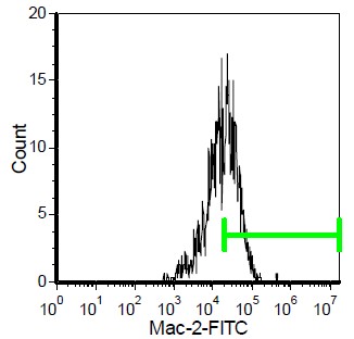
| Note | Protocol: Flow Cytometry Analysis: 1. Prepare a cell suspension in media A. For cell preparations, deplete the red blood cell population 2. Wash 2 times. 3. Resuspend the cells to a concentration of 2x10e7 cells/ml in media A. Add 50 µl of this suspension to each tube (each tube will then contain 1x10e6 cells, representing 1 test). 4. To each tube, add 0.5-1.0 µg of antibody per 10e6 cells. 5. Vortex the tubes to ensure thorough mixing of antibody and cells. 6. Incubate the tubes for 30 minutes at 4°C. 7. Wash 2 times at 4°C. 8. Add 100 µl of secondary antibody FITC Goat anti-rat IgG (H+L) at appropriate dilution. 9. Incubate the tubes at 4°C for 30-60 minutes. (It is recommended that the tubes are protected from light since most fluorochromes are light sensitive). 10. Wash 2 times at 4°C in media B. 11. Resuspend the cell pellet in 50 µl ice cold media B. 12. Transfer to suitable tubes for flow cytometric analysis containing 15 µl of propidium iodide at 0.5 mg/ml in PBS. This stains dead cells by intercalating in DNA. Media: A. Phosphate buffered saline (pH 7.2) + 5% normal serum of host species + sodium azide (100 µl of 2M sodium azide in 100 mls). B. Phosphate buffered saline (pH 7.2) + 0.5% Bovine serum albumin + sodium azide (100 µl of 2M sodium azide in 100 mls). Results of Tissue Distribution by Flow Cytometry Analysis: - Mouse Strain: BALB/c - Cell concentration: 1 x 10e6 cells per test - Antibody concentration used: 0.5 µg/10e6 cells - Isotypic Control: Rat IgG2a Percentage of cells stained above control: Thymus = 2.41% Thioglycollate-elicited peritoneal exudate = 85.8% (see picture below). |
| Reference Data | |
Documents
| Product Manuals |
| FAQs |
| SDS |
{0} Product Review(s)
0 Product Review(s)
Submit review
Be the first one to submit a review
Product Citations
*Delivery time may vary from web posted schedule. Occasional delays may occur due to unforeseen
complexities in the preparation of your product. International customers may expect an additional 1-2 weeks
in shipping.


 Germany
 Japan
Japan
 United Kingdom
 China
China


