


 Islands")


")


")


")


")
")


 Germany
 Japan
Japan
 United Kingdom
 China
China
MHC Class II RT1D Mouse Monoclonal Antibody [Clone ID: OX-17]
Product Images
Specifications
| Product Data | |
| Clone Name | OX-17 |
| Applications | FC, IHC |
| Recommended Dilution | Flow Cytometry. Immunohistochemistry on frozen sections. |
| Reactivities | Rat |
| Host | Mouse |
| Isotype | IgG1 |
| Clonality | Monoclonal |
| Immunogen | Rat spleen membrane glycoproteins depleted of Ia-A antigens. Immunocyte Donor: BALB/c spleen Fusion Partner: X63 Ag8.653 |
| Specificity | This monoclonal antibody recognizes a monomorphic determinant on the a chain of the rat Ia antigen and appears to be the rat homologue of mouse Ia-E. It recognizes the rat Ia product present on B, but not T cells from lymph node or thoracic duct lymph. It does not bind to thymocytes or erythrocytes. The antibody does not cross-react with rat Ia-A or mouse Ia-E antigen, but rabbit antibody raised against the antibody affinity column-purified MRC OX-17 antigen cross-reacted on tissues of mice expressing Ia-E mouse antigen but not on those mouse strains that were Ia-E antigen negative. |
| Formulation | PBS, 0.02% NaN3 and EIA grade BSA as a stabilizing protein to bring total protein concentration to 4-5 mg/ml Label: Biotin State: Liquid purified Ig |
| Concentration | 0.1 mg/ml |
| Purification | Protein G Chromatography |
| Conjugation | Biotin |
| Synonyms | HLA Class II |
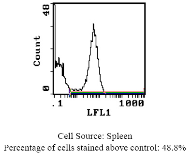
| Note | Protocol: FLOW CYTOMETRY ANALYSIS: Method: 1. Prepare a cell suspension in media A. For cell preparations, deplete the red blood cell population with Lympholyte®-Rat cell separation medium. 2. Wash 2 times. 3. Resuspend the cells to a concentration of 2x10e7 cells/ml in media A. Add 50 µl of this suspension to each tube (each tube will then contain 1 x 10e6 cells, representing 1 test). 4. To each tube, add 0.5-0.1 µg* of this Ab per 10e6 cells. 5. Vortex the tubes to ensure thorough mixing of antibody and cells. 6. Incubate the tubes for 30 minutes at 4°C. 7. Wash 2 times at 4°C. 8. Add 100 µl of secondary antibody (Streptavidin-FITC) at a 1:500 dilution. 9. Incubate tubes at 4°C for 30 - 60 minutes (It is recommended that tubes are protected from light since most fluorochromes are light sensitive). 10. Wash 2 times at 4°C. 11. Resuspend the cell pellet in 50 µl ice cold media B. 12. Transfer to suitable tubes for flow cytometric analysis containing 15 µl of propidium iodide at 0.5 mg/ml in PBS. This stains dead cells by intercalating in DNA. Media: A. Phosphate buffered saline (pH 7.2) + 5% normal serum of host species + sodium azide (100 µl of 2M sodium azide in 100 mls). B. Phosphate buffered saline (pH 7.2) + 0.5% Bovine serum albumin + sodium azide (100 µl of 2M sodium azide in 100 mls). Results - Tissue Distribution: Rat Strain: Fischer Cell Concentration: 1x10e6 cells per test Antibody Concentration Used: 0.1 µg/10e6 cells Isotypic Control: Biotin Mouse IgG1 Cell Source Percentage of cells stained above control: Thymus 6.1% Spleen 48.8% Lymph Node 27.5% |
| Reference Data | |
Documents
| Product Manuals |
| FAQs |
| SDS |
{0} Product Review(s)
0 Product Review(s)
Submit review
Be the first one to submit a review


Product Citations
*Delivery time may vary from web posted schedule. Occasional delays may occur due to unforeseen
complexities in the preparation of your product. International customers may expect an additional 1-2 weeks
in shipping.