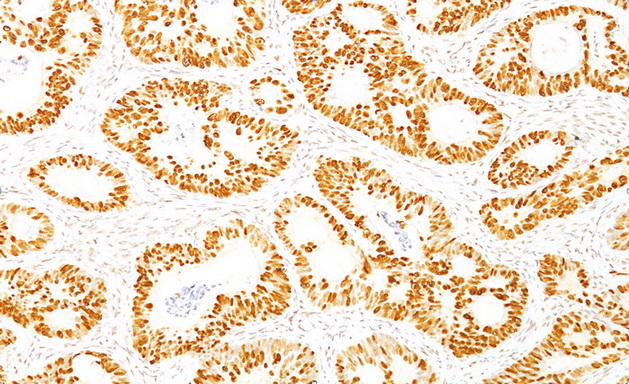
TP53 / p53 (Wild type + Mutant) Mouse Monoclonal Antibody [Clone ID: DO-7]
CAT#: SM2139PT
TP53 / p53 (Wild type + Mutant) mouse monoclonal antibody, clone DO-7, Purified
Specifications
| Product Data | |
| Clone Name | DO-7 |
| Applications | FC, IF, IHC, IP, WB |
| Recommended Dilution | ELISA: Use BSA free Antibody for Coating. Western Blot: 0.5-1 µg/ml. Flow Cytometry: 0.5-1 µg/106 cells. Immunofluorescence: 0.5-1 µg/ml. Immunoprecipitation: 0.5-1 µg/500 µg protein lysate. Immunohistochemistry on Frozen and Formalin-Fixed Paraffin Sections: 0.25-0.5 µg/ml for 30 minutes at RT. Staining of formalin-fixed tissues requires boiling tissue sections in 10mM citrate buffer, pH 6.0, for 10-20 min followed by cooling at RT for 20 minutes. Positive Control: MDA-MB-231 Cells. Breast or Colon carcinoma. |
| Reactivities | Bovine, Human, Monkey |
| Host | Mouse |
| Isotype | IgG2b |
| Clonality | Monoclonal |
| Immunogen | Recombinant Human wild type p53 protein expressed in E. coli. |
| Specificity | Recognizes a 53kDa protein, which is identified as p53 suppressor gene product. It reacts with the mutant as well as the wild form of p53. Its epitope maps within the N-terminus (aa 37-45) of p53. Monoclonal antibody PAb1801 does not block the binding of DO-7 MAb to p53 in an ELISA test. p53 is a tumor suppressor gene expressed in a wide variety of tissue types and is involved in regulating cell growth, replication, and apoptosis. It binds to MDM2, SV40 T antigen and human papilloma virus E6 protein. Positive nuclear staining with p53 antibody has been reported to be a negative prognostic factor in breast carcinoma, lung carcinoma, colorectal, and urothelial carcinoma. Anti-p53 positivity has also been used to differentiate uterine serous carcinoma from endometrioid carcinoma as well as to detect intratubular germ cell neoplasia. Mutations involving p53 are found in a wide variety of malignant tumors, including breast, ovarian, bladder, colon, lung, and melanoma. Cellular Localization: Nuclear. |
| Formulation | 10mM PBS State: Purified State: Liquid purified IgG fraction from Bioreactor Concentrate Stabilizer: 0.05% BSA Preservative: 0.05% Sodium Azide |
| Purification | Protein A/G Chromatography |
| Predicted Protein Size | 53 kDa |
| Background | p53 is a tumor suppressor gene expressed in a wide variety of tissue types and is involved in regulating cell growth, replication, and apoptosis. It binds to mdm2, SV40 T antigen and human papilloma virus E6 protein p53 senses DNA damage and possibly facilitating repair. Mutation involving p53 is found in a wide variety of malignant tumors, including breast, ovarian, bladder, colon, lung, and melanoma. |
| Synonyms | Cellular tumor antigen p53, Tumor suppressor p53, Phosphoprotein p53, NY-CO-13 |
| Reference Data | |
Documents
| Product Manuals |
| FAQs |
| SDS |
{0} Product Review(s)
0 Product Review(s)
Submit review
Be the first one to submit a review
Product Citations
*Delivery time may vary from web posted schedule. Occasional delays may occur due to unforeseen
complexities in the preparation of your product. International customers may expect an additional 1-2 weeks
in shipping.


 Germany
 Japan
Japan
 United Kingdom
 China
China


