Cd3d Mouse Monoclonal Antibody [Clone ID: 1F4]
Specifications
| Product Data | |
| Clone Name | 1F4 |
| Applications | FC, FN, IHC, IP |
| Recommended Dilution | Immunoprecipitation. Flow Cytometry: Use 10 µl of 1/10-1/25 diluted antibody to label 106 cells in 100 µl. Immunohistochemistry on Frozen Sections: 1/10-1/25. Immunohistochemistry on Paraffin Sections: 1/10. This clone is suitable for use on Paraffin Embedded material using target unmasking fluid (Ref.3). Functional Assay: Functionally the addition of the antibody to a culture of Rat T cells induces the proliferation of T-cells in the presence of PMA. We recommend the use of Azide Free SM253A for this purpose. |
| Reactivities | Rat |
| Host | Mouse |
| Isotype | IgM |
| Clonality | Monoclonal |
| Immunogen | F344 rat T cells stimulated with PMA (TPA) and calcium ionophore. |
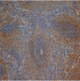
| Specificity | This antibody recognizes Rat CD3, a 25kD antigen which is found on rat T-cells. This antibody does not react with Rat B cells. In immunohistology it stains Rat thymus tissues strongly in the medulla and weakly in the cortex. |
| Formulation | PBS containing 0.09% Sodium Azide as preservative. State: Purified State: Liquid purified IgM fraction. |
| Concentration | 1.0 mg/ml |
| Purification | Ammonium Sulphate Precipitation. |
| Database Link | |
| Background | T cell activation through the antigen receptor (TCR) involves the cytoplasmic tails of the CD3 subunits: CD3 gamma, CD3 delta, CD3 epsilon and CD3 zeta. These CD3 subunits are structurally related members of the immunoglobulins super family encoded by closely linked genes on human chromosome 11. The CD3 components have long cytoplasmic tails that associate with cytoplasmic signal transduction molecules. This association is mediated at least in part by a double tyrosine based motif present in a single copy in the CD3 subunits. CD3 may play a role in TCR induced growth arrest, cell survival and proliferation. The CD3 antigen is present on 68-82% of normal peripheral blood lymphocytes, 65-85% of thymocytes and Purkinje cells in the cerebellum. It is never expressed on B or NK cells. Decreased percentages of T lymphocytes may be observed in some autoimmune diseases. |
| Synonyms | CD3D, T3D |
| Reference Data | |
Documents
| Product Manuals |
| FAQs |
| SDS |
{0} Product Review(s)
0 Product Review(s)
Submit review
Be the first one to submit a review
Product Citations
*Delivery time may vary from web posted schedule. Occasional delays may occur due to unforeseen
complexities in the preparation of your product. International customers may expect an additional 1-2 weeks
in shipping.


 Germany
 Japan
Japan
 United Kingdom
 China
China


