


 Islands")


")


")


")


")
")


 Germany
 Japan
Japan
 United Kingdom
 China
China
Icam1 Mouse Monoclonal Antibody [Clone ID: 1A29]
Product Images
Specifications
| Product Data | |
| Clone Name | 1A29 |
| Applications | FC, FN, IHC, IP |
| Recommended Dilution | Immunoprecipitation. Flow cytometry. Immunohistochemistry on frozen sections. In vivo and in vitro function blocking (1,2,3,4,5,6). |
| Reactivities | Rat |
| Host | Mouse |
| Isotype | IgG1 |
| Clonality | Monoclonal |
| Specificity | This antibody recognizes the intercellular adhesion molecule-1, designated as CD54. It inhibits homotypic aggregation of PHA blasts. Immunoprecipitation analysis shows that the antigen has features identical to those of human ICAM-1. Antigen distribution is in full agreement with that reported with the human ICAM-1. |
| Formulation | PBS, pH 7.4, 0.09 % sodium azide (NaN3) and 1 % BSA Label: Biotin State: Liquid purified Ig fraction |
| Concentration | 0.1 mg/ml |
| Conjugation | Biotin |
| Database Link | |
| Background | ICAM-1 is a 90 kDa adhesion molecule belonging to the superimmunoglobulin family. It is a cell surface ligand of the lymphocyte integrin, LFA-1 (lymphocyte function associated antigen-1) and is known to play an important role in various cell-cell interactions in the immune system. ICAM-1 exists on fibroblasts, epithelial and endothelial cells. |
| Synonyms | ICAM-1 |
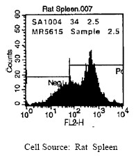
| Note | Protocol: FLOW CYTOMETRY ANALYSIS: Method: 1. Prepare a cell suspension in media A. For cell preparations, deplete the red blood cell population Rat cell separation medium. 2. Wash 2 times. 3. Resuspend the cells to a concentration of 2x10e7 cells/ml in media A. Add 50 µl of this suspension to each tube (each tube will then contain 1x10e6 cells, representing 1 test). 4. To each tube, add ~0.25 µg of antibody. 5. Vortex the tubes to ensure thorough mixing of antibody and cells. 6. Incubate the tubes for 30 minutes at 4°C. 7. Wash 2 times at 4°C. 8. Add 100 µl of detection reagent Streptavidin-PE at a concentration of 1:50. 9. Incubate the tubes at 4°C for 30-60 minutes. (It is recommended that the tubes are protected from light since most fluorochromes are light sensitive). 10. Wash 2 times at 4°C in media B. 11. Resuspend the cell pellet in 50 µl ice cold media B. 12. Transfer to suitable tubes for flow cytometric analysis containing 15 µl of propidium iodide at 0.5 mg/ml in PBS. This stains dead cells by intercalating in DNA. Media: A. Phosphate buffered saline (pH 7.2) + 5% normal serum of host species + sodium azide (100 µl of 2M sodium azide in 100 mls). B. Phosphate buffered saline (pH 7.2) + 0.5% Bovine serum albumin + sodium azide (100 µl of 2M sodium azide in 100 mls). Results: Tissue Distribution by Flow Cytometry Analysis: (Representative Histogram) Rat Strain: Wistar Cell Concentration: 1 X 10e6 cells per test Antibody Concentration Used: 0.25 μg/ 10e6 cells Isotypic Control: Strepdavidin-PE (see picture below) IMMUNOHISTOCHEMISTRY: Method: 1. Dilute the antibody 1:500 - 1:1000 to stain tissue sections. 2. Cryostat sections should be fixed in cold acetone and incubated with 50-100 µl of diluted antibody/tissue sections. |
| Reference Data | |
Documents
| Product Manuals |
| FAQs |
| SDS |
{0} Product Review(s)
0 Product Review(s)
Submit review
Be the first one to submit a review


Product Citations
*Delivery time may vary from web posted schedule. Occasional delays may occur due to unforeseen
complexities in the preparation of your product. International customers may expect an additional 1-2 weeks
in shipping.