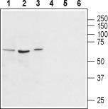
Avpr1a Rabbit Polyclonal Antibody
Other products for "Avpr1a"
Specifications
| Product Data | |
| Applications | WB |
| Recommended Dilution | WB: 1:200-1:2000; IHC: 1:100-1:3,000; FC: 1:50-1:600 |
| Reactivities | Human, Mouse, Rat |
| Host | Rabbit |
| Clonality | Polyclonal |
| Immunogen | Peptide (C)KFAKDDSDSMSRR, corresponding to amino acid residues 378-390 of rat Vasopressin V1A receptor (Accession P30560). Intracellular, C-terminus. |
| Formulation | Lyophilized. Concentration before lyophilization ~0.8mg/ml (lot dependent, please refer to CoA along with shipment for actual concentration). Buffer before lyophilization: phosphate buffered saline (PBS), pH 7.4, 1% BSA, 0.05% NaN3. |
| Purification | Affinity purified on immobilized antigen. |
| Conjugation | Unconjugated |
| Storage | Store at -20°C as received. |
| Stability | Stable for 12 months from date of receipt. |
| Gene Name | arginine vasopressin receptor 1A |
| Database Link | |
| Background | Vasopressin (AVP), the antidiuretic hormone, is a cyclic nonapeptide involved in the homeostasis of body fluid, blood volume, vascular tone, and blood pressure. AVP also belongs to the family of vasoactive and mitogenic peptides involved in physiological and pathological cell growth and differentiation1. AVP exerts its actions through binding to specific V1A, V1B, and V2, membrane receptors coupled to distinct second messengers2. V1 AVP receptor has the typical features of a G-protein coupled transmembrane receptor with seven putative hydrophobic domains, connected by three extracellular and three intracellular loops3. V1 receptors activate phospholipases A2, C, and D, resulting in the production of inositol 1,4,5-trisphosphate (IPS) and 1,Z-diacylglycerol (DAG), the mobilization of intracellular Ca2+, the influx of extracellular Ca2+, the activation of protein kinase C, and protein phosphorylation4. V1A AVP receptors have been shown by radioligand binding techniques to be present in vascular smooth muscle cells, hepatocytes, blood platelets, lymphocytes and monocytes, type 2 pneumocytes, adrenal cortex, brain (hippocampus septum et amygdalae), reproductive organs, retinal epithelium, renal mesangial cells, and the A10, A7r5,3T3, and WRK-1 cell lines4. V1A AVP receptors mediate cell contraction and proliferation, platelet aggregation, coagulation factor release, and glycogenolysis. V1B AVP receptors are located in the anterior pituitary where they stimulate ACTH release1. |
| Synonyms | AVPR1; V1aR |
| Reference Data | |
Documents
| Product Manuals |
| FAQs |
{0} Product Review(s)
0 Product Review(s)
Submit review
Be the first one to submit a review
Product Citations
*Delivery time may vary from web posted schedule. Occasional delays may occur due to unforeseen
complexities in the preparation of your product. International customers may expect an additional 1-2 weeks
in shipping.


 Germany
 Japan
Japan
 United Kingdom
 China
China


