GFAP Mouse Monoclonal Antibody [Clone ID: 5C10]
Other products for "GFAP"
Specifications
| Product Data | |
| Clone Name | 5C10 |
| Applications | IF, WB |
| Recommended Dilution | WB: 1:5000, IF: 1:500-1:1000, IHC: 1:500-1:1000, IHC-F: 1:500-1:1000, IHC-P: 1:500-1:1000 |
| Reactivities | Bovine, Chicken, Equine, Human, Mouse, Rat, Avian |
| Host | Mouse |
| Isotype | IgG1, kappa |
| Clonality | Monoclonal |
| Immunogen | A preparation of purified pig spinal cord GFAP |
| Formulation | Preservative: 0.05% Sodium Azide. Aliquot and store at -20C or -80C. Avoid freeze-thaw cycles. |
| Concentration | lot specific |
| Purification | Ascites |
| Conjugation | Unconjugated |
| Storage | Store at -20°C as received. |
| Stability | Stable for 12 months from date of receipt. |
| Predicted Protein Size | 55 kDa |
| Gene Name | glial fibrillary acidic protein |
| Database Link | |
| Background | GFAP (glial fibrillary acidic protein) is a class-III intermediate filament protein which is a cell-specific marker with ability to distinguish astrocytes from other glial cells during CNS development. Originally discovered as a major fibrous protein of multiple sclerosis plaques, GFAP was subsequently characterized as a member of intermediate filament protein family (GFAP, peripherin, desmin and vimentin). In Western blot assay, GFAP depicts ~50-55kD band which is generally associated with few lower molecule weight bands of its alternate transcripts. GFAP is especially expressed in astrocytes and certain other astroglia in CNS, in satellite cells in peripheral ganglia, and in non-myelinating Schwann cells in PNS. Astrocytes react to damage/pathologies leading to "astrogliosis" wherein the reactive astrocytes show enhanced GFAP expression and neural stem cells also express high GFAP levels. Defects in GFAP have been linked to Alexander disease (ALEXD) which is characterized by the presence of Rosenthal fibers, GFAP containing cytoplasmic inclusions in astrocytes. |
| Synonyms | ALXDRD |
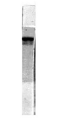
| Note | This GFAP (5C10) antibody is useful for Immunocytochemistry/Immunofluorescence, Immunohistochemistry on paraffin-embedded and frozen sections, and Western blot. In WB, a band can be seen at ~55 kDa. |
| Reference Data | |
| Protein Families | ES Cell Differentiation/IPS |
Documents
| Product Manuals |
| FAQs |
{0} Product Review(s)
0 Product Review(s)
Submit review
Be the first one to submit a review
Product Citations
*Delivery time may vary from web posted schedule. Occasional delays may occur due to unforeseen
complexities in the preparation of your product. International customers may expect an additional 1-2 weeks
in shipping.


 Germany
 Japan
Japan
 United Kingdom
 China
China


