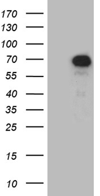
PIP5K3 (PIKFYVE) Mouse Monoclonal Antibody [Clone ID: OTI2F11]
CAT#: TA810465
PIKFYVE mouse monoclonal antibody,clone OTI2F11
Size: 30 ul
Formulation: Carrier Free
View other "OTI2F11" antibodies (2)
Special Offer: Get a 15% discount on this product. Use code: “NEURO15".
Specifications
| Product Data | |
| Clone Name | OTI2F11 |
| Applications | WB |
| Recommended Dilution | WB 1:2000 |
| Reactivities | Human, Mouse, Rat |
| Host | Mouse |
| Isotype | IgG2a |
| Clonality | Monoclonal |
| Immunogen | Full length human recombinant protein of human PIKFYVE (NP_001171471) produced in E.coli. |
| Formulation | PBS (PH 7.3) containing 1% BSA, 50% glycerol and 0.02% sodium azide. |
| Concentration | 1 mg/ml |
| Purification | Purified from mouse ascites fluids or tissue culture supernatant by affinity chromatography (protein A/G) |
| Conjugation | Unconjugated |
| Storage | Store at -20°C as received. |
| Stability | Stable for 12 months from date of receipt. |
| Gene Name | phosphoinositide kinase, FYVE-type zinc finger containing |
| Database Link | |
| Background | Phosphorylated derivatives of phosphatidylinositol (PtdIns) regulate cytoskeletal functions, membrane trafficking, and receptor signaling by recruiting protein complexes to cell- and endosomal-membranes. Humans have multiple PtdIns proteins that differ by the degree and position of phosphorylation of the inositol ring. This gene encodes an enzyme (PIKfyve; also known as phosphatidylinositol-3-phosphate 5-kinase type III or PIPKIII) that phosphorylates the D-5 position in PtdIns and phosphatidylinositol-3-phosphate (PtdIns3P) to make PtdIns5P and PtdIns(3,5)biphosphate. The D-5 position also can be phosphorylated by type I PtdIns4P-5-kinases (PIP5Ks) that are encoded by distinct genes and preferentially phosphorylate D-4 phosphorylated PtdIns. In contrast, PIKfyve preferentially phosphorylates D-3 phosphorylated PtdIns. In addition to being a lipid kinase, PIKfyve also has protein kinase activity. PIKfyve regulates endomembrane homeostasis and plays a role in the biogenesis of endosome carrier vesicles from early endosomes. Mutations in this gene cause corneal fleck dystrophy (CFD); an autosomal dominant disorder characterized by numerous small white flecks present in all layers of the corneal stroma. Histologically, these flecks appear to be keratocytes distended with lipid and mucopolysaccharide filled intracytoplasmic vacuoles. Alternative splicing results in multiple transcript variants encoding distinct isoforms. [provided by RefSeq, May 2010] |
| Synonyms | CFD; FAB1; HEL37; PIP5K; PIP5K3; ZFYVE29 |
| Reference Data | |
| Protein Families | Druggable Genome |
| Protein Pathways | Endocytosis, Fc gamma R-mediated phagocytosis, Inositol phosphate metabolism, Metabolic pathways, Phosphatidylinositol signaling system, Regulation of actin cytoskeleton |
Documents
| Product Manuals |
| FAQs |
| SDS |
Resources
| Antibody Resources |
{0} Product Review(s)
Be the first one to submit a review


 Germany
 Japan
Japan
 United Kingdom
 China
China


