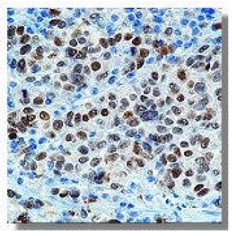
MITF Mouse Monoclonal Antibody [Clone ID: C5]
Specifications
| Product Data | |
| Clone Name | C5 |
| Applications | EMSA, IHC, IP, WB |
| Recommended Dilution | Gel Supershift. Western Blot (1 µg/ml). Immunoprecipitation (2 µg/mg of protein lysate). Immunohistochemistry on Frozen and Paraffin Sections. Positive Control: 501 Mel Human melanoma cells, wild-type Human, Rat, Mouse osteoclast cells. |
| Reactivities | Human, Mouse, Rat |
| Host | Mouse |
| Isotype | IgG1 |
| Clonality | Monoclonal |
| Immunogen | Hybridoma produced by the fusion of splenocytes from RBF/DnJ mice immunized with an N-terminal fragment of Human microphthalmia protein and mouse myeloma NS1 cells. |
| Specificity | Recognizes in Western blotting a doublet of 52-56kDa, identified as serine-phosphorylated and unphosphorylated forms of melanocytic isoforms of microphthalmia (Mi) transcription factor. This antibody does not cross-react with other b-HLH-ZIP factors by DNA mobility shift assay. |
| Formulation | PBS containing 0.08% Sodium Azide as preservative State: Purified State: Liquid purified IgG fraction |
| Concentration | lot specific |
| Purification | Protein A/G Chromatography |
| Storage | Upon receipt, store undiluted (in aliquots) at -20°C. Avoid repeated freezing and thawing. |
| Stability | Shelf life: one year from despatch. |
| Predicted Protein Size | 52-56 kDa |
| Database Link | |
| Background | There are two known isoforms of MiTF differing by 66 amino acids at the NH2 terminus. Shorter forms are expressed in melanocytes and run as two bands at 52kDa and 56kDa, while the longer Mi form runs as a cluster of bands at 60-70kDa in osteoclasts and in B16 melonoma cells (but not other melanoma cell lines), as well as mast cells and heart. It reacts with both melanocytic as well as the non- melanocytic isoforms of MiTF. Mi is a basic helix-loop-helix-leucin zipper (b-HLH-ZIP) transtripotion factor implicated in pigmentation, mast cells and bone development. The mutation of MiTF causes Waardenburg Syndrome type II in humans. In mice, a profound loss of pigmented cells in the skin eye and inner ear results, as well as osteopetrosis and defects in natural killer and mast cells. These melanocyte isoforms have been shown by two dimensional tryptic mapping to differ in c-Kit-induced phosphorylation. Osteopetrotic rat strain harbors a large genomic deletion encompassing the 3 |
| Synonyms | Microphthalmia-associated transcription factor, Mi-protein |
| Reference Data | |
Documents
| Product Manuals |
| FAQs |
{0} Product Review(s)
0 Product Review(s)
Submit review
Be the first one to submit a review
Product Citations
*Delivery time may vary from web posted schedule. Occasional delays may occur due to unforeseen
complexities in the preparation of your product. International customers may expect an additional 1-2 weeks
in shipping.


 Germany
 Japan
Japan
 United Kingdom
 China
China


