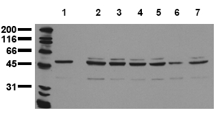
Aurora A (AURKA) (N-term) (incl. pos. control) Mouse Monoclonal Antibody [Clone ID: 7F12]
CAT#: AM20207PU-N
Aurora A (AURKA) (N-term) (incl. pos. control) mouse monoclonal antibody, clone 7F12, Purified
Other products for "AURKA"
Specifications
| Product Data | |
| Clone Name | 7F12 |
| Applications | WB |
| Recommended Dilution | Immunobotting (Western Blot): 0.5 µg/ml for HRPO/ECL detection. Recommended blocking buffer: Casein/Tween 20 based blocking buffer and blot incubation buffer. Included Postitive Control: Cell Lysate from untreated A431 cells. |
| Reactivities | Canine, Human, Mouse, Rat |
| Host | Mouse |
| Isotype | IgG1 |
| Clonality | Monoclonal |
| Immunogen | Peptide conjugated to hemocyanin Epitope: N-terminus. |
| Specificity | Recognizes Aurora-A (N-term). |
| Formulation | PBS / 0.09% Sodium Azide / PEG and Sucrose. State: Purified State: Lyophilized purified IgG fraction. |
| Reconstitution Method | Restore with 1 ml H2O (15 min, RT). |
| Purification | Size Exclusion Chromatography. |
| Storage | Store lyophilized (preferably in a desiccator) at -20°C and reconstituted (aliquote and freeze in liquid nitrogen) at -20°C to -80°C. Avoid repeated freezing and thawing. Thaw aliquots at 37°C. Thawed aliquots may be stored at 2-8°C up to 3 months. |
| Stability | Shelf life: one year from despatch. |
| Gene Name | Homo sapiens aurora kinase A (AURKA), transcript variant 2 |
| Database Link | |
| Background | Aurora proteins are members of a serine/threonine kinase family. They play a crucial role in mitosis by regulating chromosome segregation and cytokinesis. There are three forms of Aurora proteins in mammalian cells: AuroraA, B and C. Aurora-A (Aurora-2; STK6, ARK1, Aurora/IPL-1 related kinase) associates with centrosomes and microtubules during mitosis. Phosphorylation of a threonine residue within the activation loop of the catalytic domain lead to activation of Aurora-A. AuroraB (Aurora-1) is responsible for chromatin modification and histone H3 phosphporylation. |
| Synonyms | AURKA, AIK1, ARK1, AURA, BTAK, STK15, STK6, Aurora/IPL1-related kinase 1, AURORA2 |
| Note | Molecular Weight: 47 kDa Protocol: Positive Control: Cell Lysate from untreated A431 cells. Formulation: Lyophilized Cell lysate from Serum starved A431 cells. Stability: Reconstitute by addition of 200 µl H2O. After complete solubilization add 200 µl 2x SDS-PAGE sample buffer, mix and incubate at 90°C for 5 min. Application: The Positive Control lysate is recommended for Immunoblot applications. 20µl of Positive Control correspond to ca. 20.000 cells. Use 20µl/lane (mini gel) for HRPO/ECL detection of the target proteins. Storage: Aliquote and store frozen. Avoid repeated freeze/thaw cycles. Shelf life: one year from despatch. The Lyophilized cell lysates contain SDS and are not recommended for applications with native proteins such as Immunoprecipitation. |
| Reference Data | |
| Protein Families | Druggable Genome, Protein Kinase, Stem cell - Pluripotency |
| Protein Pathways | Oocyte meiosis |
Documents
| Product Manuals |
| FAQs |
{0} Product Review(s)
0 Product Review(s)
Submit review
Be the first one to submit a review
Product Citations
*Delivery time may vary from web posted schedule. Occasional delays may occur due to unforeseen
complexities in the preparation of your product. International customers may expect an additional 1-2 weeks
in shipping.


 Germany
 Japan
Japan
 United Kingdom
 China
China


