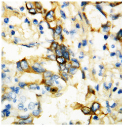
TNFRSF1A (195-211) Rabbit Polyclonal Antibody
Other products for "TNFRSF1A"
Specifications
| Product Data | |
| Applications | IHC, WB |
| Recommended Dilution | Western blot: 0.1-0.5 μg/ml. Immunohistochemistry on Paraffin Sections: 0.5-1 μg/ml. By Heat: Boiling the paraffin sections in 10mM citrate buffer, pH6.0, for 20mins is required for the staining of formalin/paraffin sections. |
| Reactivities | Goat, Human, Mouse, Rat |
| Host | Rabbit |
| Isotype | IgG |
| Clonality | Polyclonal |
| Immunogen | A synthetic peptide corresponding to a sequencein the middle region of Human TNFR1 (aa 195-211). |
| Specificity | This antibody detects CD120a / TNFR1 (195-211). No cross reactivity with other proteins. |
| Formulation | 0.9 mg NaCl, 0.2 mg Na2HPO4 State: Aff - Purified State: Lyophilized Ig fraction Stabilizer: 5 mg BSA Preservative: 0.05 mg Thimerosal, 0.05 mg Sodium Azide |
| Reconstitution Method | 0.2 ml of distilled water will yield a concentration of 0.5 mg/ml. |
| Purification | Immunoaffinity Chromatography |
| Storage | Store lyophilized at 2-8°C for 6 months or at -20°C long term. After reconstitution store the antibody undiluted at 2-8°C for one month or (in aliquots) at -20°C long term. Avoid repeated freezing and thawing. |
| Stability | Shelf life: one year from despatch. |
| Gene Name | Homo sapiens tumor necrosis factor receptor superfamily member 1A (TNFRSF1A) |
| Database Link | |
| Background | Tumor necrosis factor receptor 1(TNFR1), a potent cytokine, elicits a broad spectrum of biologic responses which are mediated by binding to a cell surface receptor. Its gene is located on 12p13.2. The coding region and the 3-prime untranslated region of TNFR1 are distributed over 10 exons. There are 2 different proteins that serve as major receptors for TNF-alpha, one associated with myeloid cells and one associated with epithelial cells. Additionally, TNFR1 associates with the MADD protein through a death domain-death domain interaction. MADD provides a physical link between TNFR1 and the induction of mitogen-activated protein (MAP) kinase (e.g., ERK2) activation and arachidonic acid release. TNFR1-induced apoptosis involves 2 sequential signaling complexes. Complex I, the initial plasma membrane-bound complex, consists of TNFR1, the adaptor TRADD, the kinase RIP1, and TRAF2 and rapidly signals activation of NF-kappa-B. In a second step, TRADD and RIP1 associate with FADD and caspase-8, forming a cytoplasmic complex, complex II. |
| Synonyms | Tumor necrosis factor receptor 1, TNF-R1, TNF-RI, TNFR-I, p55, p60, Tnfrsf1a |
| Reference Data | |
| Protein Families | Druggable Genome, Secreted Protein, Transcription Factors, Transmembrane |
| Protein Pathways | Adipocytokine signaling pathway, Alzheimer's disease, Amyotrophic lateral sclerosis (ALS), Apoptosis, Cytokine-cytokine receptor interaction, MAPK signaling pathway |
Documents
| Product Manuals |
| FAQs |
{0} Product Review(s)
0 Product Review(s)
Submit review
Be the first one to submit a review
Product Citations
*Delivery time may vary from web posted schedule. Occasional delays may occur due to unforeseen
complexities in the preparation of your product. International customers may expect an additional 1-2 weeks
in shipping.


 Germany
 Japan
Japan
 United Kingdom
 China
China


